ვინაიდან დაავადება სწრაფად პროგრესირებადია, ადრეული ამოცნობა ძალიან მნიშვნელოვანია. ნეკროზული ფასციიტი სიცოცხლისათვის საშიში და კრიტიკული ქირურგიული გადაუდებელი შემთხვევაა.[2]Sartelli M, Guirao X, Hardcastle TC, et al. 2018 WSES/SIS-E consensus conference: recommendations for the management of skin and soft-tissue infections. World J Emerg Surg. 2018 Dec 14;13:58.
https://wjes.biomedcentral.com/articles/10.1186/s13017-018-0219-9
http://www.ncbi.nlm.nih.gov/pubmed/30564282?tool=bestpractice.com
[4]Diab J, Bannan A, Pollitt T. Necrotising fasciitis. BMJ. 2020 Apr 27;369:m1428.შეიძლება განვითარდეს სეფსისი და პოლიორგანული უკმარისობა.[2]Sartelli M, Guirao X, Hardcastle TC, et al. 2018 WSES/SIS-E consensus conference: recommendations for the management of skin and soft-tissue infections. World J Emerg Surg. 2018 Dec 14;13:58.
https://wjes.biomedcentral.com/articles/10.1186/s13017-018-0219-9
http://www.ncbi.nlm.nih.gov/pubmed/30564282?tool=bestpractice.com
[34]Bonne SL, Kadri SS. Evaluation and management of necrotizing soft tissue infections. Infect Dis Clin North Am. 2017 Sep;31(3):497-511.
http://www.ncbi.nlm.nih.gov/pubmed/28779832?tool=bestpractice.com
აუცილებელია სასწრაფოდ ქირურგიულ გუნდთან მიმართვის და კრიტიკული მართვის საჭიროების შეფასების უზრუნველყოფა. ნეკროზული ფასციიტი არის ძირითადად კლინიკური დიაგნოზი, დინამიკაში სწრაფად განვითარებადი ნიშნებითა და სიმპტომებით, მიუხედავად იმისა, რომ აუცილებელია ლაბორატორიული გამოკვლევა, მიკრობიოლოგიური გამოკვლევა, და გამოსახულებითი გამოკვლევა, როდესაც ეს შესაძლებელია.[2]Sartelli M, Guirao X, Hardcastle TC, et al. 2018 WSES/SIS-E consensus conference: recommendations for the management of skin and soft-tissue infections. World J Emerg Surg. 2018 Dec 14;13:58.
https://wjes.biomedcentral.com/articles/10.1186/s13017-018-0219-9
http://www.ncbi.nlm.nih.gov/pubmed/30564282?tool=bestpractice.com
[4]Diab J, Bannan A, Pollitt T. Necrotising fasciitis. BMJ. 2020 Apr 27;369:m1428.
ანამნეზი.
საჭიროა განისაზღვროს სიმპტომების პროგრესირების დრო და სიჩქარე. რისკ-ფქტორების გამოკვლევა, როგორიცაა:
კანის დაზიანება ან მთლიანობის დარღვევა
ტრავმა, ოპერაციული ჩარევა
მანეკროზებელი ფასციიტი, რომელიც უკავშირდება ცოტა ხნის წინ გადატანილ აბდომინალურ ქირურგიას, ან ვითარდება საზარდულის არეში, მეტი ალბათობით იქნება პოლიმიკრობული.
ქრონიკული დაავადების ფონზე განვითარებული იმუნოსუპრესიული მდგომარეობა(მაგ.:შაქრიანი დიაბეტი, ალკოჰოლზე დამოკიდებულება)
ნარკოტიკების ინტრავენური გამოყენება
ჩუტყვავილა
ჰერპეს-ზოსტერი
მნიშვნელოვანია გვახსოვდეს, რომ კანის დაზიანება შეიძლება იყოს უმნიშვნელო, მცირე ზომის (მაგ.მწერის ნაკბენი) ან პაციენტს არც კი ახსოვდეს.[1]Pasternack MS, Swartz MN. Cellulitis, necrotizing fasciitis, and subcutaneous tissue infections. In: Bennett JE, Dolin R, Blaser MJ, eds. Mandell, Douglas, and Bennett’s principles and practice of infectious diseases. Philadelphia, PA: Elsevier; 2015:1194-215.[25]Stevens DL, Aldape MJ, Bryant AE. Necrotizing fasciitis, gas gangrene, myositis and myonecrosis. In: Cohen J, Powderly WG, Opal SM, eds. Infectious diseases. Amsterdam, Netherlands: Elsevier; 2017:95-103.e1.ექსპოზიციის ისტორია ზოგჯერ შეიძლება ინფორმაციული იყოს და განისაზღვროს სავარაუდო გამომწვევი (მაგ. მტკნარი წყლის ექსპოზიცია ასოცირებულია Aeeromonas hydrophila-თან , ზღვის წყალის ექსპოზიცია ან ნედლი ხამანწკების მოხმარება ასოცირებულაი Vibrio vunlificus-თან); მიუხედავად ამისა, საწყისი ემპირიულად შერჩეული ანტიბიოტიკი უნდა იყოს ფართო სპექტრის და არ უნდა იყოს დაფუძნებული მხოლოდ ანამნეზში არსებულ ექსპოზიციებზე..[9]Kuo YL, Shieh SJ, Chiu HY, et al. Necrotizing fasciitis caused by Vibrio vulnificus: epidemiology, clinical findings, treatment and prevention. Eur J Clin Microbiol Infect Dis. 2007 Nov;26(11):785-92.
http://www.ncbi.nlm.nih.gov/pubmed/17674061?tool=bestpractice.com
[15]Hua C, Urbina T, Bosc R, et al. Necrotising soft-tissue infections. Lancet Infect Dis. 2023 Mar;23(3):e81-94.
http://www.ncbi.nlm.nih.gov/pubmed/36252579?tool=bestpractice.com
[25]Stevens DL, Aldape MJ, Bryant AE. Necrotizing fasciitis, gas gangrene, myositis and myonecrosis. In: Cohen J, Powderly WG, Opal SM, eds. Infectious diseases. Amsterdam, Netherlands: Elsevier; 2017:95-103.e1.
ცელულიტის მქონე პაციენტში მანეკროზებელი ფასციიტის სხვა შესაძლო სიმპტომებია თავბრუსხვევა, გულის ფრიალი (პალპიტაციები), გულისრევა ან ღებინება ან დელირიუმი.
გასინჯვა
დიაგნოზი უნდა განვიხილოთ პაციენტში, რომელსაც აქვს ცელულუტი და კლინიკური საფრთხილო ნიშნები, როგორიცაა სეფსისთან დაკავშირებული ორგანოების უკმარისობის სწრაფი შეფასება (qSOFA; სპეციფიკურად არ არის ვალიდური ნეკროზული ფასციიტისთვის). ფსიქიკური მდგომარეობის ცვლილება (გლაზგოს კომის შკალა ≤15), სისტოლური ჰიპოტენზია (სისტოლური არტერიული წნევა ≤100 მმ.ვწყ.სვ.) და სუნთქვის სიხშირის მატება (≥22 ჩასუნთქვა წუთში) მიანიშნებს, რომ ცელულიტის მქონე პაციენტს შესაძლოა განვითარებული ჰქონდეს მანეკროზებელი პროცესი, რომელიც საჭიროებს ქირურგიულ შეფასებას.[2]Sartelli M, Guirao X, Hardcastle TC, et al. 2018 WSES/SIS-E consensus conference: recommendations for the management of skin and soft-tissue infections. World J Emerg Surg. 2018 Dec 14;13:58.
https://wjes.biomedcentral.com/articles/10.1186/s13017-018-0219-9
http://www.ncbi.nlm.nih.gov/pubmed/30564282?tool=bestpractice.com
პაციენტს შეიძლება აღენიშნებოდეს არასპეციფიკური და არაადგილობრივი სიმპტომები (მაგ., მწვავედ განვითარებული შეუძლოდ ყოფნა ნორმალური ტემპერატურის ფონზე ) ან შეიძლება გამოვლინდეს მძიმე ნიშნები პოლიორგანული დისფუნქციისა და შოკის ნიშნებით.[2]Sartelli M, Guirao X, Hardcastle TC, et al. 2018 WSES/SIS-E consensus conference: recommendations for the management of skin and soft-tissue infections. World J Emerg Surg. 2018 Dec 14;13:58.
https://wjes.biomedcentral.com/articles/10.1186/s13017-018-0219-9
http://www.ncbi.nlm.nih.gov/pubmed/30564282?tool=bestpractice.com
ცელულიტის არეში ყრუ ან ინტენსიური ტკივილის განვითარება შესაძლოა მიუთითებდეს კანქვეშა ინფექციის არსებობაზე.[1]Pasternack MS, Swartz MN. Cellulitis, necrotizing fasciitis, and subcutaneous tissue infections. In: Bennett JE, Dolin R, Blaser MJ, eds. Mandell, Douglas, and Bennett’s principles and practice of infectious diseases. Philadelphia, PA: Elsevier; 2015:1194-215.[16]Hasham S, Matteucci P, Stanley PR, et al. Necrotising fasciitis. BMJ. 2005 Apr 9;330(7495):830-3. [Erratum in: BMJ. 2005 May 14;330(7500):1143].
http://www.ncbi.nlm.nih.gov/pubmed/15817551?tool=bestpractice.com
[19]Cheung JP, Fung B, Tang WM, et al. A review of necrotising fasciitis in the extremities. Hong Kong Med J. 2009 Feb;15(1):44-52.
http://www.hkmj.org/system/files/hkm0902p44.pdf
http://www.ncbi.nlm.nih.gov/pubmed/19197096?tool=bestpractice.com
[20]Angoules AG, Kontakis G, Drakoulakis E, et al. Necrotising fasciitis of upper and lower limb: a systematic review. Injury. 2007 Dec;38(suppl 5):S19-26.
http://www.ncbi.nlm.nih.gov/pubmed/18048033?tool=bestpractice.com
[35]Endorf FW, Cancio LC, Klein MB. Necrotizing soft-tissue infections: clinical guidelines. J Burn Care Res. 2009 Sep-Oct;30(5):769-75.
http://www.ncbi.nlm.nih.gov/pubmed/19692912?tool=bestpractice.com
მანეკროზებელი ფასციიტის თანმხლები ტკივილი შესაძლებელია არ იყოს კანის ხილული ცვლილების ხარისხის პროპორციული. აუცილებელია იმის გათვალისწინება, რომ მანეკროზებელი ფასციიტის მქონე პაციენტებს შესაძლოა აღენიშნოს ზემდებარე კანის ნორმალური საფარველი და რომ A ჯგუფის სტრეპტოკოკებით გამოწვეული მანეკროზებელი ფასციიტის დროს კანის ცვლილება მოგვიანებით ნიშანს წარმოადგენს.
ცელულიტის არეში კანის შემოწმებისას ზოგ შემთხვევაში ვლინდება კრეპიტაცია, ვეზიკულები, ბულები, შეფერილობის ცვლილება ბაცი მორუხო ელფერით, ან შეშუპება სიწითლის გარეთაც.[3]Sartelli M, Coccolini F, Kluger Y, et al. WSES/GAIS/WSIS/SIS-E/AAST global clinical pathways for patients with skin and soft tissue infections. World J Emerg Surg. 2022 Jan 15;17(1):3.
https://wjes.biomedcentral.com/articles/10.1186/s13017-022-00406-2
http://www.ncbi.nlm.nih.gov/pubmed/35033131?tool=bestpractice.com
კანის შეუმჩნეველი ცვლილებები, სითხის გამოჟონვა და შეშუპება, წინ უძღვის კანის ხილული ცვლილებების, ბუშტუკებისა და სიწითლის, განვითარებას.
შემთხვევათა დაახლოებით ნახევარში ინფექციის კერა ვითარდება კიდურებზე, ხოლო დანარჩენი შემთხვევები ლოკალიზებულია შორისის, ტორსის, თავის და კისრის არეებში.[1]Pasternack MS, Swartz MN. Cellulitis, necrotizing fasciitis, and subcutaneous tissue infections. In: Bennett JE, Dolin R, Blaser MJ, eds. Mandell, Douglas, and Bennett’s principles and practice of infectious diseases. Philadelphia, PA: Elsevier; 2015:1194-215.[2]Sartelli M, Guirao X, Hardcastle TC, et al. 2018 WSES/SIS-E consensus conference: recommendations for the management of skin and soft-tissue infections. World J Emerg Surg. 2018 Dec 14;13:58.
https://wjes.biomedcentral.com/articles/10.1186/s13017-018-0219-9
http://www.ncbi.nlm.nih.gov/pubmed/30564282?tool=bestpractice.com
[3]Sartelli M, Coccolini F, Kluger Y, et al. WSES/GAIS/WSIS/SIS-E/AAST global clinical pathways for patients with skin and soft tissue infections. World J Emerg Surg. 2022 Jan 15;17(1):3.
https://wjes.biomedcentral.com/articles/10.1186/s13017-022-00406-2
http://www.ncbi.nlm.nih.gov/pubmed/35033131?tool=bestpractice.com
[4]Diab J, Bannan A, Pollitt T. Necrotising fasciitis. BMJ. 2020 Apr 27;369:m1428.[16]Hasham S, Matteucci P, Stanley PR, et al. Necrotising fasciitis. BMJ. 2005 Apr 9;330(7495):830-3. [Erratum in: BMJ. 2005 May 14;330(7500):1143].
http://www.ncbi.nlm.nih.gov/pubmed/15817551?tool=bestpractice.com
[19]Cheung JP, Fung B, Tang WM, et al. A review of necrotising fasciitis in the extremities. Hong Kong Med J. 2009 Feb;15(1):44-52.
http://www.hkmj.org/system/files/hkm0902p44.pdf
http://www.ncbi.nlm.nih.gov/pubmed/19197096?tool=bestpractice.com
[20]Angoules AG, Kontakis G, Drakoulakis E, et al. Necrotising fasciitis of upper and lower limb: a systematic review. Injury. 2007 Dec;38(suppl 5):S19-26.
http://www.ncbi.nlm.nih.gov/pubmed/18048033?tool=bestpractice.com
ყველაზე გავრცელებული ლოკალიზაცია A ჯგუფის სტრეპტოკოკებით გამოწვეული ნეკროზული ფასციიტისთვის არის ბარძაყი და კიდურები. კიდურზე, განსაკუთრებით ზედა კიდურზე განვითარებული ნეკროზული ფასციიტი მეტი ალბათობით უკავშირდება A ჯგუფის სტრეპტოკოკებს და არა პოლიმიკრობულ ინფექციას. ნეკროზული ფასციიტის ზოგიერთ შემთხვევას შესაძლოა თან ახლდეს მასთან ასოცირებული მიოზიტი, მომიჯნავე კუნთებზე პროცესის გავრცელების გამო. ეს უფრო დამახასიათებელია A ჯგუფის სტრეპტოკოკული, და არა პოლიმიკრობული ინფექციებისთვის.
მანეკროზებელი ფასციიტი, რომელიც უკავშირდება ცოტა ხნის წინ გადატანილ აბდომინალურ ქირურგიას, ან ვითარდება საზარდულის არეში, მეტი ალბათობით იქნება პოლიმიკრობული.
ლაბორატორიიული კვლევები
სავარაუდოდ ნეკროზული ფასციიტით ჰოსპიტალიზებულ ყველა პაციენტს სასწრაფოდ ესაჭიროება სისხლის საერთო ანალიზი ლეიკოციტური ფორმულით, შარდოვანას, ელექტროლიტების, კრეატინინის და C-რეაქტიული ცილის (CRP) განსაზღვრა. ამ ბიომარკერებიდან ზოგიერთი პროგნოზული ქულების მისაღებად გამოიყენება. მათ შორის ნეკროზული ფასციიტის ლაბორატორიული რისკის ინდიკატორის (LRINEC). ლაქტატისა და პროკალციტონინის მატება შეიძლება ასევე დაკავშირებული იყოს ავადობისა და სიკვდილიანობის გაზრდილ ალბათობასთან.[15]Hua C, Urbina T, Bosc R, et al. Necrotising soft-tissue infections. Lancet Infect Dis. 2023 Mar;23(3):e81-94.
http://www.ncbi.nlm.nih.gov/pubmed/36252579?tool=bestpractice.com
რესპირატორულ დარღვევებზე ეჭვის შემთხვევაში საჭიროა გაკეთდეს არტერიული სისხლის აირების კონტროლი.
მანეკროზებელი ფასციტი ხშირად ასოცირებულია ლაბორატორული მონაცემების სხვადასხვა არა-სპეციფიურ ცვლილებებთან, კერძოდ:
ლეიკოციტების (WBC) პათოლოგიურად მაღალ ან დაბალ რაოდენობასთან მარცხნივ გადახრით, ან მის გარეშე (პოლიმორფონუკლეარული ლეიკოციტების და/ან უმწიფარი ფორმების მომატებული პროცენტული მაჩვენებელი). ლეიკოციტების (WBC) დაბალი რაოდენობა შესაძლოა მიანიშნებდეს მძიმე ფორმის სეფსისის არსებობაზე
შარდოვანას და კრეატინინის დონეების მომატებასთან ინტრაცელულარული (უჯრედშიდა) მოცულობის შემცირების შედეგად
შრატში ნატრიუმის შემცველობის შემცირებასთან
C-რეაქტიული პროტეინის (CRP) მატებასთან
შრატში კრეატინ კინაზას მომატება
პლაზმაში ლაქტატის მატება.
გამომწვევის იდენტიფიცირების მიზნით სისხლის ნიმუშის აღება კულტივირებისათვის საჭიროა ანტიბიოტიკოთერაპიის დაწყებამდე.[15]Hua C, Urbina T, Bosc R, et al. Necrotising soft-tissue infections. Lancet Infect Dis. 2023 Mar;23(3):e81-94.
http://www.ncbi.nlm.nih.gov/pubmed/36252579?tool=bestpractice.com
ბაქტერიოლოგიური დიაგნოზის დასმის მიზნით ასევე გამოიყენება ასპირაციული მასალა, ბიოფსიით მიღებული ან ქირურგიული ჩარევის დროს აღებული ქსოვილოვანი მასალა. გრამის წესით შეღებვისა და კულტივირების მიზნით.[5]Stevens DL, Bisno AL, Chambers HF, et al. Practice guidelines for the diagnosis and management of skin and soft tissue infections: 2014 update by the Infectious Diseases Society of America. Clin Infect Dis. 2014 Jul 15;59(2):e10-52.
https://academic.oup.com/cid/article/59/2/e10/2895845
http://www.ncbi.nlm.nih.gov/pubmed/24973422?tool=bestpractice.com
[15]Hua C, Urbina T, Bosc R, et al. Necrotising soft-tissue infections. Lancet Infect Dis. 2023 Mar;23(3):e81-94.
http://www.ncbi.nlm.nih.gov/pubmed/36252579?tool=bestpractice.com
თავიდანვე უნდა იყოს აღებული კანის ნაცხი.[15]Hua C, Urbina T, Bosc R, et al. Necrotising soft-tissue infections. Lancet Infect Dis. 2023 Mar;23(3):e81-94.
http://www.ncbi.nlm.nih.gov/pubmed/36252579?tool=bestpractice.com
მოლეკულური ტესტირებით შესაძლებელია დამატებითი პათოგენების იდენტიფიცირება.[15]Hua C, Urbina T, Bosc R, et al. Necrotising soft-tissue infections. Lancet Infect Dis. 2023 Mar;23(3):e81-94.
http://www.ncbi.nlm.nih.gov/pubmed/36252579?tool=bestpractice.com
გამოსახულების მიღება
როდესაც, კლინიკურად საეჭვოა ნეკროზული ფასციიტის დიაგნოზი, რადიოლოგიური გამოკვლევების გამო არ უნდა დაყოვნდეს და გადავადდეს საჭირო ქირურგიული ჩარევა.[2]Sartelli M, Guirao X, Hardcastle TC, et al. 2018 WSES/SIS-E consensus conference: recommendations for the management of skin and soft-tissue infections. World J Emerg Surg. 2018 Dec 14;13:58.
https://wjes.biomedcentral.com/articles/10.1186/s13017-018-0219-9
http://www.ncbi.nlm.nih.gov/pubmed/30564282?tool=bestpractice.com
[3]Sartelli M, Coccolini F, Kluger Y, et al. WSES/GAIS/WSIS/SIS-E/AAST global clinical pathways for patients with skin and soft tissue infections. World J Emerg Surg. 2022 Jan 15;17(1):3.
https://wjes.biomedcentral.com/articles/10.1186/s13017-022-00406-2
http://www.ncbi.nlm.nih.gov/pubmed/35033131?tool=bestpractice.com
[5]Stevens DL, Bisno AL, Chambers HF, et al. Practice guidelines for the diagnosis and management of skin and soft tissue infections: 2014 update by the Infectious Diseases Society of America. Clin Infect Dis. 2014 Jul 15;59(2):e10-52.
https://academic.oup.com/cid/article/59/2/e10/2895845
http://www.ncbi.nlm.nih.gov/pubmed/24973422?tool=bestpractice.com
[15]Hua C, Urbina T, Bosc R, et al. Necrotising soft-tissue infections. Lancet Infect Dis. 2023 Mar;23(3):e81-94.
http://www.ncbi.nlm.nih.gov/pubmed/36252579?tool=bestpractice.com
კლინიკურად სტაბილურ პაციენტებში რადიოლოგიური კვლევით შესაძლებელია მანეკროზებელი პროცესის ნიშნების გამოვლენა.
დავადების ადრეულ სტადიაზე გამოსახვითი გამოკვლევით პათოლოგია არ ვლინდება, დაავადების პროგრესირებასთან ერთად ,მოგვიანებით სტადიებზე ,შესაძლებელია გამოჩნდეს კანქვეშა აირები. რბილ ქსოვილებში აირების ვიზუალიზაცია შეიძლება მიუთითებდეს რბილი ქსოვილების პათოლოგიურ ცვლილებებზე დ ამყარებს ეჭვს დაგნოზზე..[1]Pasternack MS, Swartz MN. Cellulitis, necrotizing fasciitis, and subcutaneous tissue infections. In: Bennett JE, Dolin R, Blaser MJ, eds. Mandell, Douglas, and Bennett’s principles and practice of infectious diseases. Philadelphia, PA: Elsevier; 2015:1194-215.[2]Sartelli M, Guirao X, Hardcastle TC, et al. 2018 WSES/SIS-E consensus conference: recommendations for the management of skin and soft-tissue infections. World J Emerg Surg. 2018 Dec 14;13:58.
https://wjes.biomedcentral.com/articles/10.1186/s13017-018-0219-9
http://www.ncbi.nlm.nih.gov/pubmed/30564282?tool=bestpractice.com
[16]Hasham S, Matteucci P, Stanley PR, et al. Necrotising fasciitis. BMJ. 2005 Apr 9;330(7495):830-3. [Erratum in: BMJ. 2005 May 14;330(7500):1143].
http://www.ncbi.nlm.nih.gov/pubmed/15817551?tool=bestpractice.com
[36]American College of Radiology. ACR appropriateness criteria: suspected osteomyelitis, septic arthritis, or soft tissue infection (excluding spine and diabetic foot). 2022 [internet publication].
https://acsearch.acr.org/docs/3094201/Narrative
თუმცა, უნდა გვახსოვდეს, რომ რბილ ქსოვილებში აირების ვიზუალიზაცია გვიანი ნიშანია და ჩვეულებრივი რენტგენოგრაფიული გამოკვლევის მგრძნობელობა ნეკროზული ინფექციის გამოსავლენად დაბალია.[15]Hua C, Urbina T, Bosc R, et al. Necrotising soft-tissue infections. Lancet Infect Dis. 2023 Mar;23(3):e81-94.
http://www.ncbi.nlm.nih.gov/pubmed/36252579?tool=bestpractice.com
ულტრაბგერითი გამოკვლევა უპირატესია. ის შეიძლება ჩატარდეს უმოკლეს დროში ავადმყოფის საწოლთან და შეიძლება დაგვეხმაროს მარტივი ცელულიტის და ნეკროზული ფასციიტის დიფერენცირებაში.[2]Sartelli M, Guirao X, Hardcastle TC, et al. 2018 WSES/SIS-E consensus conference: recommendations for the management of skin and soft-tissue infections. World J Emerg Surg. 2018 Dec 14;13:58.
https://wjes.biomedcentral.com/articles/10.1186/s13017-018-0219-9
http://www.ncbi.nlm.nih.gov/pubmed/30564282?tool=bestpractice.com
ერთი პროსპექტული კვლევის თანახმად, ულტრაბგერის შედეგები ან კანქვეშა ქსოვილის დიფუზური გასქელება, რომელსაც სითხის დაგროვება ახლავს თან 4 მმ-ზე ღრმად, არის 88% სენსიტიური და 93% სპეციფიკური ნიშანი.[37]Yen ZS, Wang HP, Ma HM, et al. Ultrasonographic screening of clinically-suspected necrotizing fasciitis. Acad Emerg Med. 2002 Dec;9(12):1448-51.
https://onlinelibrary.wiley.com/doi/abs/10.1197/aemj.9.12.1448?sid=nlm%3Apubmed
http://www.ncbi.nlm.nih.gov/pubmed/12460854?tool=bestpractice.com
კომპიუტერული ტომოგრაფია და მაგნიტურ რეზონანსული ტომოგრაფია მაღალსენსიტიური მეთოდებია.[3]Sartelli M, Coccolini F, Kluger Y, et al. WSES/GAIS/WSIS/SIS-E/AAST global clinical pathways for patients with skin and soft tissue infections. World J Emerg Surg. 2022 Jan 15;17(1):3.
https://wjes.biomedcentral.com/articles/10.1186/s13017-022-00406-2
http://www.ncbi.nlm.nih.gov/pubmed/35033131?tool=bestpractice.com
[36]American College of Radiology. ACR appropriateness criteria: suspected osteomyelitis, septic arthritis, or soft tissue infection (excluding spine and diabetic foot). 2022 [internet publication].
https://acsearch.acr.org/docs/3094201/Narrative
CT ,უპირატესად ,ნაჩვენებია მუცლის ღრუს , კისრისა და სახის ინფექციების დროს, ინფექციის შეჭრის კერისა და ქირურგიული ჩარევის განსაზღვრის მიზნით. კიდურების ან პერიფერიული რბილი ქსოვილების ნეკროზული ინფექციის დიაგნოსტიკაში CT ნაკლებად მნიშვნელოვანია.[15]Hua C, Urbina T, Bosc R, et al. Necrotising soft-tissue infections. Lancet Infect Dis. 2023 Mar;23(3):e81-94.
http://www.ncbi.nlm.nih.gov/pubmed/36252579?tool=bestpractice.com
[36]American College of Radiology. ACR appropriateness criteria: suspected osteomyelitis, septic arthritis, or soft tissue infection (excluding spine and diabetic foot). 2022 [internet publication].
https://acsearch.acr.org/docs/3094201/Narrative
კიდურების რბილი ქსოვილების ნეკროზული ინფექციების შესაფასებლად უფრო მგრძნობიარეა MRI, შეიძლება გამოვლინდეს ფასციის გასქელება, ღრმა ფასციალური სითხე ან შეშუპება თუმცა, ეს ნიშნები არ არის სპეციფიკური და შეიძლება გამოვლინდეს რბილი ქსოვილების სხვა ინფექციების დროსაც, მაგ. როგორიცაა ცელულიტი. გარდა ამისა,MRI -ის ჩატარების ორგანიზება შეიძლება რთული იყოს გადაუდებელ შემთხვევებში. ამდენად MRI არ ითვლება პირველი რიგის სადიაგნოსტიკო საშუალებად.[2]Sartelli M, Guirao X, Hardcastle TC, et al. 2018 WSES/SIS-E consensus conference: recommendations for the management of skin and soft-tissue infections. World J Emerg Surg. 2018 Dec 14;13:58.
https://wjes.biomedcentral.com/articles/10.1186/s13017-018-0219-9
http://www.ncbi.nlm.nih.gov/pubmed/30564282?tool=bestpractice.com
[3]Sartelli M, Coccolini F, Kluger Y, et al. WSES/GAIS/WSIS/SIS-E/AAST global clinical pathways for patients with skin and soft tissue infections. World J Emerg Surg. 2022 Jan 15;17(1):3.
https://wjes.biomedcentral.com/articles/10.1186/s13017-022-00406-2
http://www.ncbi.nlm.nih.gov/pubmed/35033131?tool=bestpractice.com
[15]Hua C, Urbina T, Bosc R, et al. Necrotising soft-tissue infections. Lancet Infect Dis. 2023 Mar;23(3):e81-94.
http://www.ncbi.nlm.nih.gov/pubmed/36252579?tool=bestpractice.com
[38]Gelbard RB, Ferrada P, Yeh DD, et al. Optimal timing of initial debridement for necrotizing soft tissue infection: a practice management guideline from the Eastern Association for the Surgery of Trauma. J Trauma Acute Care Surg. 2018 Jul;85(1):208-14.
http://www.ncbi.nlm.nih.gov/pubmed/29485428?tool=bestpractice.com
რბილი ქსოვილების ნეკროზული ინფექციის დიაგნოსტიკის მეტა-ანალიზით გამოვლინდა: CT-ს მგრძნობელობა 88.5%, სპეციფიურობა 93.3 ; მარტივი რენტგენოგრაფიის მგრძნობელობა 48.9% ,სპეციფიურობა 94.0%.[39]Fernando SM, Tran A, Cheng W, et al. Necrotizing soft tissue infection: diagnostic accuracy of physical examination, imaging, and LRINEC Score: a systematic review and meta-analysis. Ann Surg. 2019 Jan;269(1):58-65.
http://www.ncbi.nlm.nih.gov/pubmed/29672405?tool=bestpractice.com
თუ დიაგნოზი გაურკვეველია კლინიკურად ან რადიოლოგიურად, რბილი ქსოვილის ბიოფსიით შეიძლება საბოლოო დიაგნოზის დადასტურება.[2]Sartelli M, Guirao X, Hardcastle TC, et al. 2018 WSES/SIS-E consensus conference: recommendations for the management of skin and soft-tissue infections. World J Emerg Surg. 2018 Dec 14;13:58.
https://wjes.biomedcentral.com/articles/10.1186/s13017-018-0219-9
http://www.ncbi.nlm.nih.gov/pubmed/30564282?tool=bestpractice.com
თუმცა, ბიოფსია გარკვეულ სირთულებთანაა დაკავშირებული. მოითხოვს მაღალკვალიფიციურ სპეციალისტს, ესაჭიროება დრო და არ არის ხელმისაწვდომი ყველა რეგიონში , მათ შორის დიდ ბრიტანეთშიც.,[2]Sartelli M, Guirao X, Hardcastle TC, et al. 2018 WSES/SIS-E consensus conference: recommendations for the management of skin and soft-tissue infections. World J Emerg Surg. 2018 Dec 14;13:58.
https://wjes.biomedcentral.com/articles/10.1186/s13017-018-0219-9
http://www.ncbi.nlm.nih.gov/pubmed/30564282?tool=bestpractice.com
დიაგნოსტიკური კრიტერიუმი
სიმძიმის შეფასების ქულების გამოყენება შეიძლება დაგეხმაროთ სისტემური სეფსისის მაღალი რისკის მქონე პაციენტებში, რომელთაც სჭირდებათ ურგენტული მომსახურება და ქირურგის კონსულტაცია.[2]Sartelli M, Guirao X, Hardcastle TC, et al. 2018 WSES/SIS-E consensus conference: recommendations for the management of skin and soft-tissue infections. World J Emerg Surg. 2018 Dec 14;13:58.
https://wjes.biomedcentral.com/articles/10.1186/s13017-018-0219-9
http://www.ncbi.nlm.nih.gov/pubmed/30564282?tool=bestpractice.com
პაციენტებში, რომელთაც აქვთ ცელულიტი და საფრთხილო კლინიკური ნიშნები , როგორიცაა სეფსისთან დაკავშირებული ორგანოების უკმარისობის გამოვლინებები,( სწრაფი შეფასებით qSOFA) უნდა იყოს განხილული ნეკროზული ფასციიტის დიაგნოზი. ორგანოების უკმარისობის სწრაფი შეფასება- qSOFA გამოიყენება ინტენსიური მკურნალობის განყოფილების ფარგლებს გარეთ. ორგანოების უკმარისობის თანმიმდევრული შეფასება -SOFA გამოიყენება ინტენსიური მკურნალობის განყოფილებაში. არ არის ჩატარებული კვლევა ნეკროზული ფასციიტის დროს. სპეციფიკურად qSOFA ან SOFA- ს გამოყენების შესახებ.
ნეკროზული ფასციიტის რისკის ლაბორატორიული ინდიკატორი (LRINEC) ქულათა სისტემაა, რომელიც ლაბორატორიული მაჩვენებლების საშუალებით გამოითვლება (CRP, ჰემოგლობინის ნატრიუმი, კრეატინინი, გლუკოზა. სისტემა გვეხმარება ნეკროზული ფასციიტისა და კანის/რბილი ქსოვილების სხვა უფრო მსუბუქი ინფექციების დიფერენცირებაში.[3]Sartelli M, Coccolini F, Kluger Y, et al. WSES/GAIS/WSIS/SIS-E/AAST global clinical pathways for patients with skin and soft tissue infections. World J Emerg Surg. 2022 Jan 15;17(1):3.
https://wjes.biomedcentral.com/articles/10.1186/s13017-022-00406-2
http://www.ncbi.nlm.nih.gov/pubmed/35033131?tool=bestpractice.com
[40]Wong CH, Khin LW, Heng KS, et al. The LRINEC (Laboratory Risk Indicator for Necrotizing Fasciitis) score: a tool for distinguishing necrotizing fasciitis from other soft tissue infections. Crit Care Med. 2004 Jul;32(7):1535-41.
http://www.ncbi.nlm.nih.gov/pubmed/15241098?tool=bestpractice.com
რბილი ქსოვილების ინფექციების დიაგნოსტირების 2019 წლის მეტა-ანალიზით გამოვლინდა LRINEC 6-ს მგრძნობელობა- 68.2% (95% CI 51.4% - 81.3%), სპეციფიურობა 84.8% (95% CI 75.8% - დან 90.9%).[39]Fernando SM, Tran A, Cheng W, et al. Necrotizing soft tissue infection: diagnostic accuracy of physical examination, imaging, and LRINEC Score: a systematic review and meta-analysis. Ann Surg. 2019 Jan;269(1):58-65.
http://www.ncbi.nlm.nih.gov/pubmed/29672405?tool=bestpractice.com
მიუხედავად იმისა, რომ ვალიდაციის კვლევებმა ვერ აჩვენა საკმარისი მგრძნობელობა ან სპეციფიურობა ნეკროზული ფასციიტის დიაგნოსტიკაში LRINEC-ის მაღალი ქულა ასოცირებულია სიკვდილიანობის მაღალ რისკთან.[39]Fernando SM, Tran A, Cheng W, et al. Necrotizing soft tissue infection: diagnostic accuracy of physical examination, imaging, and LRINEC Score: a systematic review and meta-analysis. Ann Surg. 2019 Jan;269(1):58-65.
http://www.ncbi.nlm.nih.gov/pubmed/29672405?tool=bestpractice.com
[41]Hsiao CT, Chang CP, Huang TY, et al. Prospective validation of the Laboratory Risk Indicator for Necrotizing Fasciitis (LRINEC) Score for necrotizing fasciitis of the extremities. PLoS One. 2020;15(1):e0227748.
https://journals.plos.org/plosone/article?id=10.1371/journal.pone.0227748
http://www.ncbi.nlm.nih.gov/pubmed/31978094?tool=bestpractice.com
[42]Hoesl V, Kempa S, Prantl L, et al. The LRINEC score-an indicator for the course and prognosis of necrotizing fasciitis? J Clin Med. 2022 Jun 22;11(13):3583.
https://www.mdpi.com/2077-0383/11/13/3583
http://www.ncbi.nlm.nih.gov/pubmed/35806870?tool=bestpractice.com
კონსულტაცია
ამ მდგომარეობაზე მაღალი ეჭვის დროს მულტიდისციპლინური მიდგომის მქონე ექსპერტების ჩართვამ შეიძლება მნიშვნელოვნად გააუმჯობესოს მენეჯმენტი და პროგნოზი,[15]Hua C, Urbina T, Bosc R, et al. Necrotising soft-tissue infections. Lancet Infect Dis. 2023 Mar;23(3):e81-94.
http://www.ncbi.nlm.nih.gov/pubmed/36252579?tool=bestpractice.com
იმის გათვალისწინებით ,რომ დაავადების ადრეული ნიშნები მწირია და არასპეციფიური, დაბალია მათი მგრძნობელობა და არ არსებობს ოპტიმალური არაინვაზიური დიაგნოსტიკური საშუალებები , პასუხისმგებელი გადაწყვეტილების მიმღები პირები უნდა ჩაერთონ რაც შეიძლება ადრეულ ეტაპზე.[15]Hua C, Urbina T, Bosc R, et al. Necrotising soft-tissue infections. Lancet Infect Dis. 2023 Mar;23(3):e81-94.
http://www.ncbi.nlm.nih.gov/pubmed/36252579?tool=bestpractice.com
ნეკროზულ ფასციიტზე ეჭვის მიტანისთანავე საჭიროა გადაუდებელი ქირურგიული კონსულტაცია ინფიცირებული ქსოვილის ინსპექციის, გამოკვლევისა და დებრიდინგისთვის (ნეკროზული, ინფიცირებული ქსოვილის მოცილება)[3]Sartelli M, Coccolini F, Kluger Y, et al. WSES/GAIS/WSIS/SIS-E/AAST global clinical pathways for patients with skin and soft tissue infections. World J Emerg Surg. 2022 Jan 15;17(1):3.
https://wjes.biomedcentral.com/articles/10.1186/s13017-022-00406-2
http://www.ncbi.nlm.nih.gov/pubmed/35033131?tool=bestpractice.com
დამატებითი ადრეული კონსულტაციები მოიცავს ინტენსიური თერაპიის კონსულტანტის ჩართვას[15]Hua C, Urbina T, Bosc R, et al. Necrotising soft-tissue infections. Lancet Infect Dis. 2023 Mar;23(3):e81-94.
http://www.ncbi.nlm.nih.gov/pubmed/36252579?tool=bestpractice.com
დასკვნითი ბაქტერიოლოგიური დიაგნოზისათვის საუკეთესო ქსოვილოვანი ნიმუშების მიღება შესაძლებელია ქირურგიული ნეკრექტომიის შედეგად.[5]Stevens DL, Bisno AL, Chambers HF, et al. Practice guidelines for the diagnosis and management of skin and soft tissue infections: 2014 update by the Infectious Diseases Society of America. Clin Infect Dis. 2014 Jul 15;59(2):e10-52.
https://academic.oup.com/cid/article/59/2/e10/2895845
http://www.ncbi.nlm.nih.gov/pubmed/24973422?tool=bestpractice.com
კლინიკურად დაზიანებული ქსოვილის გრამის წესით შეღებვა გვაძლევს გამომწვევი მიკროორგანიზმის (ების) ადრეული გამოვლენის საშუალებას. მაგალითად, გრამ-დადებითი კოკების მოკლე ჯაჭვები მიგვანიშნებენ სტრეპტოკოკულ ინფექციაზე; კოკების დიდი ზომის გროვები მიუთითებს Staphylococcus aureus არსებობაზე. ქსოვილოვანი ნიმუშებიდან ან სისხლიდან მიღებული კულტურა უზრუნველყოფს ბაქტერიული გამომწვევის იდენტიფიცირებას. ინფექცია შესაძლოა იყოს მონომიკრობული, ან პოლიმიკრობული.[1]Pasternack MS, Swartz MN. Cellulitis, necrotizing fasciitis, and subcutaneous tissue infections. In: Bennett JE, Dolin R, Blaser MJ, eds. Mandell, Douglas, and Bennett’s principles and practice of infectious diseases. Philadelphia, PA: Elsevier; 2015:1194-215.[5]Stevens DL, Bisno AL, Chambers HF, et al. Practice guidelines for the diagnosis and management of skin and soft tissue infections: 2014 update by the Infectious Diseases Society of America. Clin Infect Dis. 2014 Jul 15;59(2):e10-52.
https://academic.oup.com/cid/article/59/2/e10/2895845
http://www.ncbi.nlm.nih.gov/pubmed/24973422?tool=bestpractice.com
[16]Hasham S, Matteucci P, Stanley PR, et al. Necrotising fasciitis. BMJ. 2005 Apr 9;330(7495):830-3. [Erratum in: BMJ. 2005 May 14;330(7500):1143].
http://www.ncbi.nlm.nih.gov/pubmed/15817551?tool=bestpractice.com
[19]Cheung JP, Fung B, Tang WM, et al. A review of necrotising fasciitis in the extremities. Hong Kong Med J. 2009 Feb;15(1):44-52.
http://www.hkmj.org/system/files/hkm0902p44.pdf
http://www.ncbi.nlm.nih.gov/pubmed/19197096?tool=bestpractice.com
[20]Angoules AG, Kontakis G, Drakoulakis E, et al. Necrotising fasciitis of upper and lower limb: a systematic review. Injury. 2007 Dec;38(suppl 5):S19-26.
http://www.ncbi.nlm.nih.gov/pubmed/18048033?tool=bestpractice.com
[Figure caption and citation for the preceding image starts]: მანეკროზებელი ფასციტის გვიანი ნიშნები გავრცობილი ცელულიტის, გამკვრივების, კანის ნეკროზის თანხლებით და ჰემორაგიული ბულების ფორმირებითFrom: Hasham S, Matteucci P, Stanley PRW, et al. Necrotising fasciitis. BMJ. 2005 Apr 9;330(7495):830-3 [Citation ends]. [Figure caption and citation for the preceding image starts]: კანის ფენობრივი გადანერგვა (STSG) ქირურგიული ნეკრექტომიის შემდეგFrom: Hasham S, Matteucci P, Stanley PRW, et al. Necrotising fasciitis. BMJ. 2005 Apr 9;330(7495):830-3 [Citation ends].
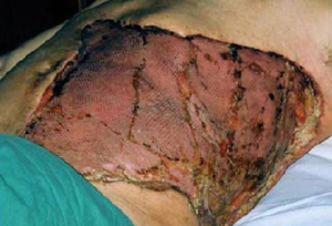
[Figure caption and citation for the preceding image starts]: მანეკროზებელი ფასციტი მუცლის მარჯვენა ნახევარში, რომელიც განუვითარდა 2 წლის გოგონას Varicella-სეული ინფექციის კვალდაკვალFrom: de Benedictis FM, Osimani P. Necrotising fasciitis complicating varicella. BMJ Case Rep. 2009;2009:bcr2008141994 [Citation ends].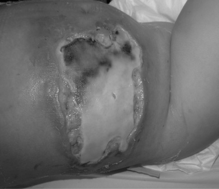 [Figure caption and citation for the preceding image starts]: კანის ნეკროზის მცირე უბნები მუცლის ქვედა მესამედის არეში ცელულიტის და მანეკროზებელი ფასციტის მქონე ახალგაზრდა ქალის შემთხვევაში, საკეისრო კვეთის გაკეთებიდან 5 დღის შემდეგFrom: Hasham S, Matteucci P, Stanley PRW, et al. Necrotising fasciitis. BMJ. 2005 Apr 9;330(7495):830-3 [Citation ends].
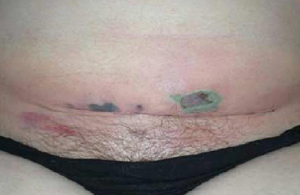
რეკომენდებულია ინფექციონისტთან კონსულტაცია ემპირიული ანტიბიოტიკების შესარჩევად, თერაპიის საჭიროებისამებრ დე-ესკალაციის მიზნით.[15]Hua C, Urbina T, Bosc R, et al. Necrotising soft-tissue infections. Lancet Infect Dis. 2023 Mar;23(3):e81-94.
http://www.ncbi.nlm.nih.gov/pubmed/36252579?tool=bestpractice.com