Images and videos
Images


Bladder cancer
European Organisation for Research and Treatment of Cancer (EORTC) risk tables: probability of recurrence and progression according to score
From World J Urol. 2007 Jun;25(3):285-95; used with permission
See this image in context in the following section/s:

Bladder cancer
Carcinoma in situ of the bladder; this can appear as a rough, erythematous patch in the bladder, but often the urothelium appears normal; random biopsy or biopsy of areas stained by 0.2% methylene blue, illustrated here, is needed to make the diagnosis
From the collection of Donald Lamm, MD, FACS
See this image in context in the following section/s:

Bladder cancer
Low-grade urothelial carcinoma seeding in the prostatic urethra; illustrated is the loop electrode used to resect bladder tumors
From the collection of Donald Lamm, MD, FACS
See this image in context in the following section/s:
Bladder cancer
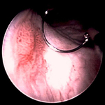
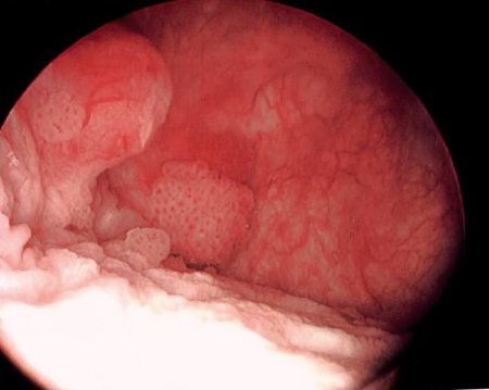
Low-grade tumors surrounded by small satellite tumors with small, uniform fronds. In the foreground is a solid tumor on a broad base, a typical appearance of high-grade tumors. Low- and high-grade tumors often occur in the same patient
From the collection of Donald Lamm, MD, FACS
See this image in context in the following section/s:
Bladder cancer
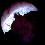
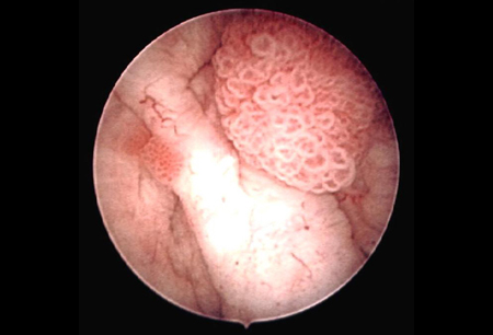
Low-grade, noninvasive (Ta) papillary urothelial carcinoma. Note adjacent satellite tumor, illustrating the field effect
From the collection of Donald Lamm, MD, FACS
See this image in context in the following section/s:

Bladder cancer
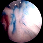
Methylene blue-stained tumor at the right bladder neck. Staining with 0.2% methylene blue (or use of the now Food and Drug Administration-approved hexaminolevulinate blue-light fluorescence cystoscopy) can help identify tumors not seen otherwise
From the collection of Donald Lamm, MD, FACS
See this image in context in the following section/s:
Bladder cancer
European Organisation for Research and Treatment of Cancer (EORTC) scoring system: weighting used to calculate recurrence and progression scores. CIS, carcinoma in situ
From World J Urol. 2007 Jun;25(3):285-95; used with permission
See this image in context in the following section/s:
Use of this content is subject to our disclaimer