Images and videos
Images


Barrett's oesophagus
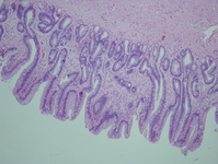
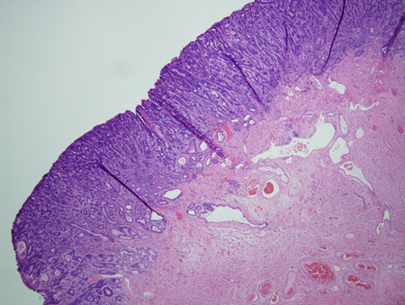
Barrett's metaplasia without dysplasia, demonstrating columnar epithelium with goblet cells from superior to the gastro-oesophageal junction
Courtesy of Adrian Ormsby, MD, Henry Ford Hospital, Detroit, MI
See this image in context in the following section/s:
Barrett's oesophagus
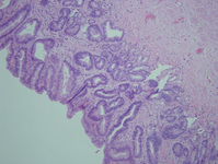
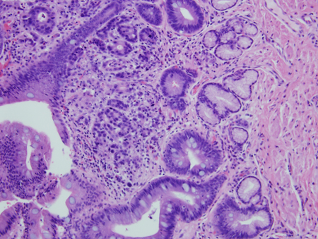
Barrett's metaplasia with high-grade dysplasia; note more advanced irregularity of the cells
Courtesy of Adrian Ormsby, MD, Henry Ford Hospital, Detroit, MI
See this image in context in the following section/s:
Barrett's oesophagus
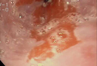
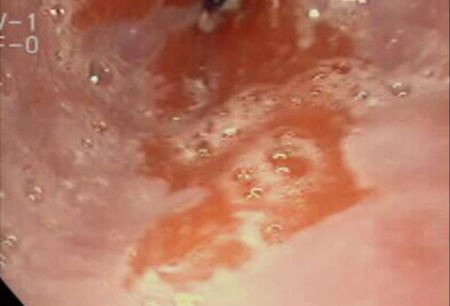
Barrett's oesophagus; note salmon-coloured mucosa extending superior to the gastro-oesophageal junction with marked irregular border
From the personal collection of Dr Vic Velanovich; used with permission
See this image in context in the following section/s:
Barrett's oesophagus
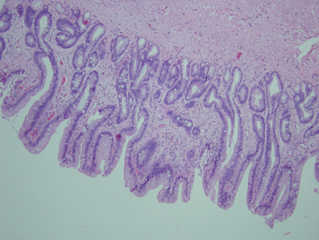
Barrett's metaplasia with high-grade dysplasia associated with a focus of intramucosal carcinoma; note the frankly malignant cells beyond the confines of the basement membrane to involve the lamina propria
Courtesy of Adrian Ormsby, MD, Henry Ford Hospital, Detroit, MI
See this image in context in the following section/s:
Barrett's oesophagus
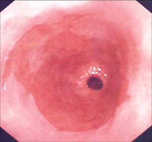
Barrett's oesophagus; note salmon-coloured mucosa extending superior to the gastro-oesophageal junction as a continuous column
From the personal collection of Dr Vic Velanovich; used with permission
See this image in context in the following section/s:

Barrett's oesophagus
Barrett's metaplasia with low-grade dysplasia; note the more irregular cells and nuclei
Courtesy of Adrian Ormsby, MD, Henry Ford Hospital, Detroit, MI
See this image in context in the following section/s:
Use of this content is subject to our disclaimer