Investigations
1st investigations to order
x-ray limb
Test
Order plain x-rays in all patients with suspected limb fracture.
Include at least two 90° orthogonal views.
Include visualisation of the joint proximal and distal to the area of suspected injury.
X-rays have limited sensitivity in early stress fractures.
If you suspect a femoral stress fracture, refer the patient for urgent x-rays of the hip and proximal femur.
[Figure caption and citation for the preceding image starts]: Radiographs showing dislocated radial head with distal third bone forearm fracturesPeter VK, Emerg Med J 2002;19:88-9; used with permission [Citation ends].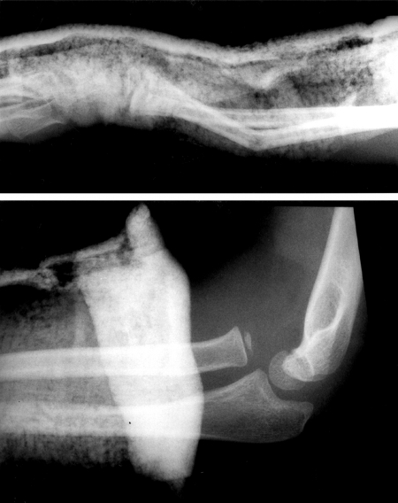 [Figure caption and citation for the preceding image starts]: X-ray showing a segmental fracture of the tibia and fibula [Citation ends].
Result
fracture and associated joint disruption
FBC, blood typing, and cross-matching (major trauma)
Test
May indicate massive bleeding, especially in cases of acute femoral fractures.
Perform as part of the initial evaluation in a major trauma setting.
Check platelets, clotting, and haemoglobin.
Result
may show acute drop in haematocrit and haemoglobin
Investigations to consider
whole body CT (adults)
Test
In adults with multiple injuries, and complex or open fractures, use a whole-body CT (including a vertex to toes scanogram followed by a vertex to mid-thigh CT scan).[51][52]
Use the findings to direct further CT of the limbs as needed.[52]
Do not routinely use whole-body CT in children under 16 years.[52]
Result
confirms fracture details and site
non-contrast CT of fracture
Test
Excellent depiction of bony anatomy. Helps to determine severity and exact nature of injury if not seen well on plain x-rays; may be useful for preoperative planning.
Three-dimensional CT reconstruction has been advocated as a way to better visualise the exact anatomy.[61]
Result
fracture details
MRI limb
Test
Excellent sensitivity and specificity for stress fractures, without the ionising radiation of a bone scan. Can also be used for definitive diagnosis of more subtle fractures.
Result
fracture line, disruption of bony architecture and/or articular surface, associated soft-tissue injury
compartment pressure testing
Test
If the examination is equivocal or diagnosis is unclear, there is a role for pressure measurement.[52][75]
May be of value in patients with impaired level of consciousness (intubated on intensive care) in whom there is an index of suspicion of compartment syndrome. However, pressure should not be relied on to rule out acute compartment syndrome if the clinical picture is consistent with a compartment syndrome. See Compartment syndrome of extremities.
If there are clinical concerns for compartment syndrome, involve orthopaedic surgeon and consider early fasciotomy.
Result
elevated pressures in acute compartment syndrome
ultrasound duplex scanning
Test
Used to assess for vascular injury, and often utilised prior to angiography.
Performed after x-rays, but must be done quickly if vascular injury is suspected.
Result
thrombosis, impaired flow at site of vascular injury
angiography
Test
Confirms clinical suspicion of vascular injury if CT angiogram is not available.
Performed after x-rays but must be done quickly if vascular injury is suspected.
Result
thrombosis, extravasation, impaired flow at site of vascular injury
dual-energy x-ray absorptiometry bone density scan
Test
Patients suspected of having osteopenia/osteoporosis should undergo bone mineral density evaluation. However, if the dual-energy x-ray absorptiometry (DXA) scan measures the femoral neck after bony healing has occurred, a falsely normal or even high bone mineral density may be reported. Thus, the contralateral femoral neck should be used for the DXA if possible.
Result
bone density low in osteoporosis/osteopenia
triple-phase bone scan
Test
Useful for early detection of stress fractures, but no longer considered a main imaging option.
If the clinical presentation is suspicious for a stress fracture but the x-rays are negative, a bone scan can be obtained. If the bone scan reveals focal, increased uptake at the area of suspicion, this is consistent with a stress fracture.
Result
focal area of increased uptake
myeloma screen
Test
If the fracture is unexplained, consider a diagnosis of myeloma and perform FBC, calcium, plasma viscosity or erythrocyte sedimentation rate, and serum and urine electrophoresis. If serum free light chains (sFLC) testing is not available, use a Bence-Jones test to check for free light chains contained in urine.[60] See Multiple Myeloma.
Result
FBC: may show anaemia; calcium may be raised; plasma viscosity or erythrocyte sedimentation rate may be raised; paraprotein identified on serum electrophoresis; sFLC or urinary Bence Jones detected
plasma viscosity or erythrocyte sedimentation rate
Test
If the fracture is unexplained, consider a diagnosis of myeloma.[60]
Result
may be raised
Use of this content is subject to our disclaimer