Images and videos
Images


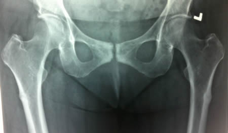
Long bone fracture
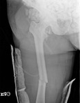
Bilateral insufficiency lesions in proximal femora in a 63-year-old woman taking weekly alendronate
BMJ Case Reports 2013 [doi:10.1136/bcr-2013-201931] Copyright © 2013 by the BMJ Publishing Group Ltd.
See this image in context in the following section/s:

Long bone fracture
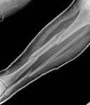
X-ray showing midshaft femur fracture
From the personal collection of Dr Philip H. Cohen
See this image in context in the following section/s:

Long bone fracture
X-ray showing osteoporotic ulnar fracture involving the proximal third of the shaft with associated dislocation of the radial head at the elbow (Monteggia fracture)
From the personal collection of Dr Philip H. Cohen
See this image in context in the following section/s:

Long bone fracture
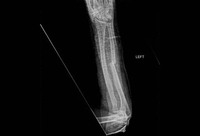
X-ray showing a segmental fracture of the tibia and fibula
From the personal collection of Dr Philip H. Cohen
See this image in context in the following section/s:
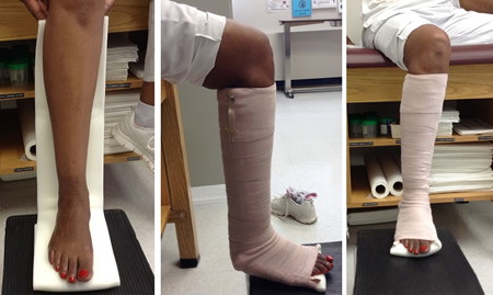
Long bone fracture
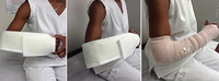
Posterior leg splint
Author (Philip Cohen)
See this image in context in the following section/s:
Long bone fracture
BMJ Rapid Recommendations: low-intensity pulsed ultrasound (LIPUS) for bone healing
Poolman RW, et al. BMJ 2017;356:j576
See this image in context in the following section/s:
Long bone fracture
OTA classification of radius and ulna fractures - locations, types, and groups
BMJ Evidence Centre
See this image in context in the following section/s:

Long bone fracture
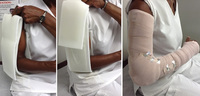
Double sugar-tong splint
Author (Philip Cohen)
See this image in context in the following section/s:
Long bone fracture
Recommended immobilization techniques for long bone fractures
Created by the BMJ Evidence Centre
See this image in context in the following section/s:

Long bone fracture
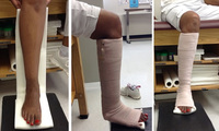
Sugar-tong splint
Author (Philip Cohen)
See this image in context in the following section/s:
Use of this content is subject to our disclaimer