Aetiology
Among otherwise healthy adults, high-energy trauma (e.g., motor vehicle accidents), sport injuries, falls, and assaults are the main causative factors.
Risk factors for insufficiency fractures include osteoporosis and other chronic metabolic disease, advanced age, prolonged corticosteroid use, female sex, lower body mass index, history of a recent fall, and prior fracture.[21] Risk factors for development of femoral stress fractures include high running mileage, female sex, and female athlete triad (osteopenia/osteoporosis, eating-disordered behaviour, and oligo/amenorrhoea).[22]
Humeral shaft fractures:
Commonly result from direct trauma to the humerus and falls onto the outstretched hand.[23][24] Less commonly, extreme muscle contraction, electrocution injury, or seizure may lead to humeral shaft fracture.[25]
Proximal humeral shaft fractures:
Typically seen in older people after a fall on the outstretched hand. Direct trauma and seizures may also lead to these fractures.[26]
Humeral stress fractures:
Primarily occur as a result of overuse among throwing athletes. Gymnasts, weightlifters, and other athletes who place repetitive high-impact or -torque loads on the humerus have also been known to sustain these injuries.
Radial and ulnar shaft fractures:
Radial shaft fractures usually result from a fall onto the outstretched or pronated wrist, or from a direct blow.[27] High-torque forces from twisting injuries can also cause such injuries.
Isolated fractures of the mid shaft of the ulna, often called nightstick fractures, usually result from a person trying to ward off a blow from a heavy, blunt object (e.g., a night stick or truncheon). If the ulnar fracture involves the proximal third of the shaft, there may be associated dislocation of the radial head at the elbow (Monteggia fracture/dislocation). Monteggia fractures are usually due to a fall onto the outstretched hand, with the elbow extended and pronated.[28]
Concomitant fractures of both the radius and the ulna are usually the result of high-energy trauma from a blow, fall, or motor vehicle accident.
Radial and ulnar stress fractures:
Primarily occur among athletes who repetitively load the bones with high forces (e.g., gymnasts).[29][30]
Femoral shaft fractures:
Generally caused by high-energy trauma, such as a motor vehicle accident, or fall from a height.
Spiral fractures of the femoral shaft may occur as a result of a twisting injury.
Comminuted and open fractures may occur from gunshot wounds or other forms of high-energy penetrating trauma.
Femoral stress fractures:
Commonly seen in athletes and runners.
Tibial and fibular shaft fractures:
Tibial shaft fractures may result from direct trauma (usually causes transverse or comminuted fractures) or indirect twisting forces (usually causes spiral or oblique fractures).[31]
High-energy trauma may result in simultaneous fracture of both the tibial and fibular shafts.
Isolated fibular fractures are typically caused by a direct blow to the outer aspect of the leg or from an external rotation force at the ankle.
Tibial and fibular stress fractures:
Pathophysiology
In otherwise healthy bone, if resorption exceeds formation, the bone will begin to wear down. There are two varieties of stress fracture: fatigue fractures and insufficiency fractures. Fatigue fractures result from repetitive submaximal stress on previously healthy bone, leading to local accelerated bone remodelling (e.g., in the setting of new or repetitive athletic activity).[1][2] Insufficiency fractures occur due to normal activity on bones that are deficient in microstructure and/or mineralisation (e.g., osteoporosis).[1][34] If a fracture develops without significant trauma in an area of diseased bone (i.e., bone that is compromised by tumour or other disease process), this is termed a pathological fracture.[35][36]
Acute fracture of a long bone causes moderate to severe pain, as well as impairment of function. Many long bone fractures are associated with significant bleeding from the bone itself and from injured soft tissues and nearby vessels. Although this bleeding can be severe and even life-threatening (e.g., when a major vessel is lacerated), the haematoma that forms around the ends of the fracture serves as the start of the fracture healing process. Acute inflammatory mediators and cells head to and proliferate in and about the haematoma. While this may lead to increased local swelling and pain, the inflammatory response is an important part of healing.
The fracture haematoma also serves as a scaffold for subsequent callus formation. Within 8 hours of injury, increased cell division occurs in the periosteum and throughout the injured bone. Over the next few days, this increased cell division becomes more localised to the area of the fracture, and this continues for several weeks. In addition, mesenchymal stem cells are recruited to the fracture site, where they multiply and differentiate into osteogenic cells. This leads to the development of a soft cartilaginous callus between the fracture ends and superficial to the periosteum. This occurs about 7 to 10 days after the injury and starts to stabilise the fracture. Simultaneously, a central, hard callus starts to form subjacent to the periosteum, between the ends of the fracture.
Multiple growth factors allow for the development and ingrowth of new blood vessels to provide an adequate blood source for the ongoing work of healing. Hard cartilaginous callus formation peaks by around day 14 post injury, and this cartilage starts to become replaced by woven bone. This remodelling process starts 3 to 4 weeks post injury, and is implemented by osteoclasts resorbing the hard callus and osteoblasts depositing lamellar bone. Eventually, the external aspect of the callus becomes lamellar bone and the internal aspect is re-formed into a medullary cavity.[37]
Assuming proper stabilisation and adequate blood supply, some long bone fractures achieve union within 8 weeks, although others (e.g., certain tibial shaft fractures) may take 4 to 6 months to heal.[38][39][40] Functional recovery of long bone fractures may take months beyond the point at which clinical and radiographic union occur, and the process of remodelling may not be fully complete for years.
Classification
General characterisation of long bone fractures
Acute
Caused by a sudden overload of forces on healthy bone that exceeds the capacity of the bone to withstand those forces.
Stress fracture
Fatigue
Insufficiency
Force load on bone is normal but fracture occurs in bone that has abnormally low density due to a non-malignant process, such as osteoporosis.[1]
Pathological fracture
A fracture that occurs in an area of diseased bone (e.g., where the bone has been weakened by a tumour or cyst).
Anatomical location of fracture
Intra-articular
Fracture line extends within a joint.
Extra-articular
Fracture does not extend into a joint; may involve the epiphysis, metaphysis, or shaft (diaphysis) of the bone. This topic focuses on extra-articular fractures.
Classification system for specific fractures[3]
Long bone fractures in adults can be categorised in several ways:
Gross anatomical location
Intra-articular versus extra-articular (head, metaphysis, or shaft)
Direction of fracture line: transverse (horizontal), oblique, spiral
Linear, stellate, comminuted
Closed versus open (soft tissue and skin overlying bone has been opened)
Impacted (when one fragment has been driven, or impacted, into another fragment) versus non-impacted
Displaced versus non-displaced
Rotated versus non-rotated
Angulated versus non-angulated
Complete versus incomplete (rare in adults)
Neer classification system for proximal humerus fractures[4]
Several classification systems exist for proximal humerus fractures, with the Neer system being the most widely used. The fracture is classified by involvement and displacement of the following 4 structural segments:
Greater tuberosity
Lesser tuberosity
Humeral head
Humeral shaft
Although many fracture lines may be seen, if no displacement is present (defined as <1 cm of separation and <45° of angulation), it is considered a 1-part fracture. If only 1 segment is displaced, a 2-part fracture is present. If 2 segments are displaced, a 3-part fracture is present. If all 4 segments are displaced, a 4-part fracture is present.
Newer classification systems have added articular surface orientation and other fracture characteristics.[5][6]
The AO/ASIF classification system of humeral shaft fractures[7]
The Arbeitsgemeinschaft fuer Osteosynthesefragen/Association for the Study of Internal Fixation (AO/ASIF) system (which was adopted and modified by the Orthopaedic Trauma Association) classifies humeral shaft fractures into three main types, each of which is further classified by specific fracture pattern.
The three main types are:
Simple (non-comminuted)
Butterfly
Comminuted
Orthopaedic Trauma Association (OTA) classification of radius and ulna fractures based on location and various features[8]
The OTA classification described fractures of the radius and ulna by location, type, group, subgroups, and qualifications. The locations, types, and groups are as follows:[Figure caption and citation for the preceding image starts]: OTA classification of radius and ulna fractures - locations, types, and groups [Citation ends].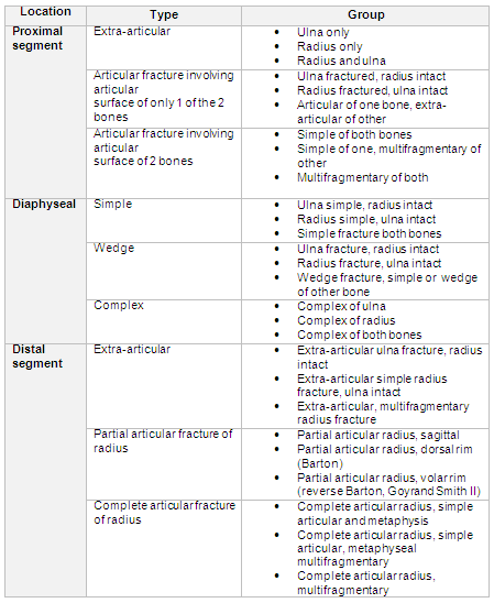
Winquist-Hansen classification system for femoral fractures[9]
Type 0 fracture is a simple transverse or oblique fracture with no comminution.
Type I fracture has a small butterfly fragment with minimal to no comminution.
Type II fracture is a butterfly fracture with ≥50% of the circumference of the 2 main fragments intact.
Type III fracture has comminution of >50% of the circumference of the major fragments.
Type IV fracture has a segmental comminution with complete loss of cortical contact.[10]
The AO Foundation classification system of tibial shaft fractures[11]
The AO Foundation classification system organises fractures into simple, wedge, or complex fractures. Each fracture is then further classified as below.
Simple fractures:
A1: spiral
A2: oblique (>30 degrees)
A3: transverse (<30 degrees)
Wedge fractures:
B1: spiral wedge
B2: bending wedge
B3: fragmented wedge
Complex fractures:
C1: spiral
C2: segmental
C3: irregular
Gustilo-Anderson classification[12]
The Gustilo and Anderson classification system organises open injuries into three categories, based on wound size, level of contamination, and osseous injury as follows:
Type I: open fracture with a clean wound less than 1 cm long.
Type II: open fracture with a laceration greater than 1 cm long without extensive soft-tissue damage, flaps, or avulsions.
Type III: open segmental fracture, open fracture with extensive soft-tissue damage, or a traumatic amputation. There are subcategories within type III for gunshot injuries, farming injuries, and any open fracture with accompanying vascular injury requiring repair.
AO Pediatric Comprehensive Classification of Long Bone Fractures (PCCF)[13]
Considers growth as a component when classifying paediatric fractures.
Use of this content is subject to our disclaimer