Images and videos
Images
Frontotemporal dementia
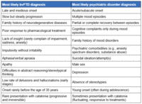
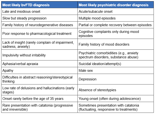
Clinical features aiding the differential diagnosis between behavioural variant FTD (bvFTD) and primary psychiatric illness
Antonioni A et al. Ex Rev of Neuro 2025; 25(3): 323-57; used with permission of Informa UK Limited, trading as Taylor & Francis Group, www.tandfonline.com
See this image in context in the following section/s:
Frontotemporal dementia
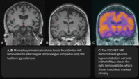
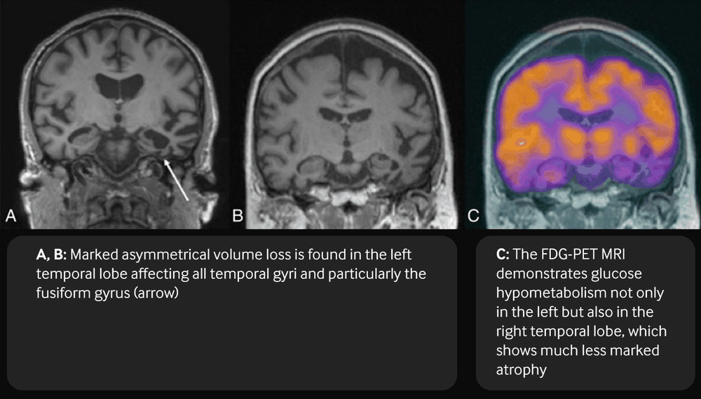
Coronal T1-weighted MRI (a, b) and fused FDG-PET MRI (c) in a patient with semantic dementia
Bhogal P et al. Eur Radiol 23, 3405-17 (2013); used with permission
See this image in context in the following section/s:
Frontotemporal dementia
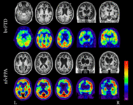
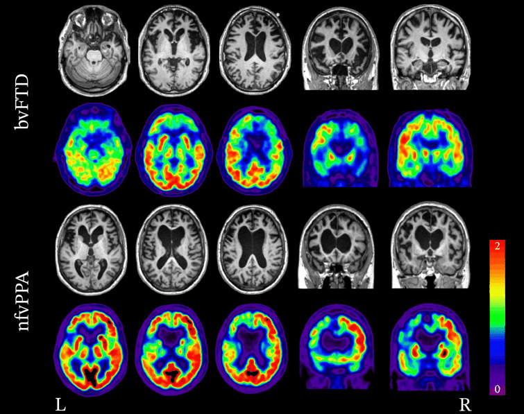
Neuroimaging patterns associated with behavioural variant FTD (bvFTD) and nonfluent variant primary progressive aphasia (nfvPPA). Structural MRI and FDG-PET demonstrating the variability in patterns of atrophy and hypometabolism in FTD. In the case of bvFTD, significant bilateral frontal lobe atrophy and hypometabolism is seen. In the case of nfvPPA, atrophy and hypometabolism is lateralised and is greatly impacting the left frontal lobe more so than the right.
Peet BT et al. Neurotherapeutics 2021 Apr; 18 (2): 728-52; used with permission
See this image in context in the following section/s:
Use of this content is subject to our disclaimer
