Investigations
1st investigations to order
x-rays
Test
Ottawa knee rules recommend x-rays in acute knee injury in an adult if any of the following features is present:[55][56]
Inability to bear weight at time of injury and at time of evaluation
Isolated tenderness at patella or fibular head
Active flexion of knee <90 degrees
Age >55.
Ottawa guidelines have excellent sensitivity (up to 100%) for detecting fractures around the knee and significantly decrease unnecessary use of x-rays. However, most acute knee injuries, including anterior cruciate ligament (ACL) tears, are not associated with fracture visible on x-ray.[64]
The absence of bony injury on x-ray does not exclude ACL injury.
Result
typically normal but may show: effusion (common); impaction fracture of the lateral femoral condyle and fracture of the posterior aspect of the lateral tibial plateau (uncommon); radiographic drawer sign (anterior subluxation of tibia on femur) (uncommon); bony avulsion of ACL at intercondylar tubercle (mainly in skeletally immature patients); mild depression on the lateral anterior femoral condyle (sulcus terminalis) contour on lateral radiograph (uncommon); lateral capsular sign (Segond's fracture), a small capsular avulsion off the lateral aspect of the proximal tibia visible on anteroposterior radiographs (uncommon but virtually pathognomic for ACL tear)
MRI
Test
MRI is the imaging modality of choice to assess for anterior cruciate ligament (ACL) injury.[59] MRI has excellent sensitivity and specificity for ACL tears, and can reveal associated injuries, such as meniscal injuries that may require additional preoperative planning and repair. False-negative MRI may occur if ACL appears grossly intact but fibres have been overstretched and are unable to function properly.[Figure caption and citation for the preceding image starts]: T1-weighted MRI showing ACL tearFrom the personal collection of Philip H. Cohen [Citation ends].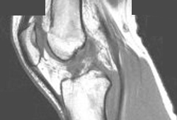 [Figure caption and citation for the preceding image starts]: T2-weighted MRI showing lateral compartment femoral condyle and tibial plateau impaction fracture, effusion, and flattening of sulcus terminalis which occurs with pivot mechanism with an ACL injury.From the personal collection of Philip H. Cohen [Citation ends].
Result
ACL fibres appear disrupted, loss of structural detail on T1-weighted images, with abnormal high signal on T2-weighted images
Use of this content is subject to our disclaimer