Approach
The key to diagnosis of an anterior cruciate ligament (ACL) injury is a thorough history and physical examination.
History
Key historical features that may indicate an ACL injury has occurred include a high-risk mechanism (contact or non-contact), a palpable or audible pop at the time of injury, rapid development of haemarthrosis, inability to return to play, pain, and a feeling that the knee is unstable or buckles with attempted weight bearing.[44] Injuries most often occur during a cutting or pivoting activity. Alternative common diagnoses in a knee injury with acute haemarthrosis include meniscus injury, osteochondral injury and fracture, and patellar dislocation.
History of prior ACL injury and female sex should also heighten suspicion.[11][12][13][14][15][31] Patients are often adolescents and young adults, or middle-aged individuals who are involved in extensive exercise.
Physical examination
Physical examination usually reveals a haemarthrosis if the injury was recent. Chronic ACL tears may or may not have an associated effusion. Acutely, range of motion is usually impaired. This may be due to a combination of pain and stiffness from haemarthrosis, as well as associated bone bruise, meniscal tears, or articular cartilage injury.[51] Tenderness is found at the lateral femoral condyle and lateral tibial plateau.[Figure caption and citation for the preceding image starts]: T1-weighted MRI showing ACL tearFrom the personal collection of Philip H. Cohen [Citation ends].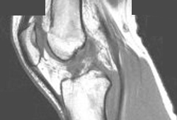
It is crucial to perform tests gently and smoothly, and to have the patient relax; otherwise the patient's discomfort and fear may lead to guarding, tensing of the hamstrings, and false-negative or inconclusive results.
Lachman's test
Lachman's test is the most accurate manoeuvre for detecting an acute ACL tear, whereas the pivot shift test performs better in chronic tears or under anaesthesia.[44][52][53] Lachman's test is performed with the patient in a supine position. The patient's knee is positioned at 20 to 30 degrees of flexion. One hand is placed on the patient's thigh and the other behind the tibia, with the clinician's thumb on the tibial tuberosity and the femur held in a stable position. The tibia is then pulled anteriorly. If the ACL is intact, a firm endpoint is felt. If this does not occur, and there is an increase in laxity compared with the uninjured knee, the test is positive, suggesting a torn ACL.[Figure caption and citation for the preceding image starts]: Lachman's manoeuvreFrom the personal collection of Philip H. Cohen [Citation ends].
Anterior drawer test
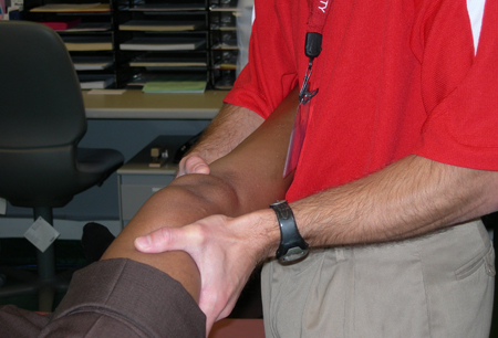
The anterior drawer test will often be positive, but is less sensitive and specific.[54] This test involves placing the patient in a supine position and flexing the hips to 45 degrees, with the knees at 90 degrees and the patient's feet on the table. Sitting on the patient's feet, the clinician takes hold of the tibia and pulls it anteriorly. If the tibia moves more than usual, the test is positive.[Figure caption and citation for the preceding image starts]: Anterior and posterior drawer test starting positionFrom the personal collection of Philip H. Cohen [Citation ends].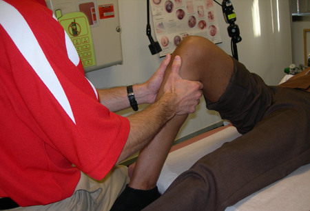
Other tests for concomitant injuries
Although not specific for identifying ACL tears, the following are necessary to evaluate for concomitant injuries that may accompany an ACL injury:
Posterior drawer test (to rule out a posterior collateral ligament sprain)
Varus/valgus stress test (to rule out a collateral ligament injury)
McMurray/Appley’s (to rule out a meniscal tear)[Figure caption and citation for the preceding image starts]: Varus and valgus stability testingFrom the personal collection of Philip H. Cohen [Citation ends].
Pivot shift manoeuvre
The pivot shift manoeuvre evaluates rotatory instability that may accompany acute ACL injuries. It may be difficult to perform and to elicit a positive test after an acute injury secondary to patient guarding from the knee injury. It is easier to perform after the acute injury pain has subsided or in the operating room under anaesthesia when patients are fully relaxed. The test is performed with the hip slightly abducted, knee fully extended, and tibia internally rotated. This would sublux the lateral tibial plateau anteriorly in an ACL deficient knee. An axial load is then applied to the knee as the knee is flexed. A palpable shift may occur when the lateral tibial plateau relocates posteriorly as the knee is flexed past 30 degrees. Whereas the Lachman‛s examination determines anteroposterior laxity, the pivot shift examination evaluates rotatory instability that may accompany acute ACL injuries.[Figure caption and citation for the preceding image starts]: Pivot startFrom the personal collection of Philip H. Cohen [Citation ends].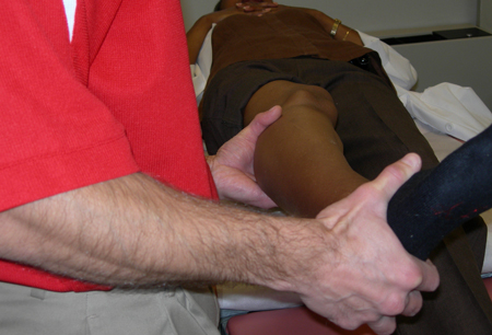 [Figure caption and citation for the preceding image starts]: Pivot finishFrom the personal collection of Philip H. Cohen [Citation ends].
Imaging
Ottawa knee rules recommend x-rays in acute knee injury in an adult if any of the following features is present:[55][56]
Inability to bear weight at time of injury and at time of evaluation
Isolated tenderness at patella or fibular head
Active flexion of knee <90 degrees
Age >55 years.
X-rays should also be performed if the clinician feels they are indicated following their assessment of the patient.
Radiographs of ACL injuries are typically negative, but may reveal a mild depression on the lateral anterior femoral condyle (sulcus terminalis) contour on a lateral knee radiograph, which may elevate the suspicion for an ACL injury pivot mechanism.[57] A lateral capsular sign (Segond's fracture), a small capsular avulsion off the lateral aspect of the proximal tibia, may also be seen on anterior to posterior radiographs.[58] This is uncommon but virtually pathognomonic for ACL tear.
Magnetic resonance imaging (MRI)
Although patients will likely have radiographs, MRI is the imaging modality of choice to assess for ACL injury.[59] MRI has excellent sensitivity and specificity for ACL tears, and can reveal associated injuries, such as meniscal injuries that may require additional preoperative planning and repair.[60][Figure caption and citation for the preceding image starts]: T1-weighted MRI showing ACL tearFrom the personal collection of Philip H. Cohen [Citation ends].[Figure caption and citation for the preceding image starts]: T2-weighted MRI showing lateral compartment femoral condyle and tibial plateau impaction fracture, effusion, and flattening of sulcus terminalis which occurs with pivot mechanism with an ACL injury.From the personal collection of Philip H. Cohen [Citation ends].
MRI is also useful when the clinical examination is impaired by factors such as patient guarding, tense effusion, or a locked joint (intra-articular block from displaced meniscal tear, etc.). MRI can also be helpful when evaluating the possibility of a torn ACL graft.[61]
From a surgical perspective, there is some evidence that preoperative MRI may be helpful in determining the suitability of a given patient's patellar tendon for use in an ACL bone-patellar tendon-bone autograft.[62]
Use of this content is subject to our disclaimer