History and exam
Key diagnostic factors
common
presence of risk factors
Key risk factors include previous groin injury, higher level of play, reduced hip abductor and adductor strength, and lower levels of training.
acute pain related to trauma
May include bony and soft-tissue injury.
history of sports-related or overuse injury
Mechanism of injury if known, level of physical activity, and/or recent changes to training programme should be discerned. Muscle strains and tendonitis frequently occur.
More common in endurance athletes or military recruits. [Figure caption and citation for the preceding image starts]: MRI demonstrating inferior right femoral neck stress fracture (compression-sided)From the collection of Cedric J. Ortiguera, MD [Citation ends].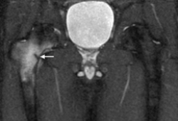
positive anterior impingement test (FADIR test)
Non-specific provocation test to elicit pain with maximal flexion, adduction, and internal rotation (FADIR) of the hip. Can be a sign of intra-articular pathology but also of several extra-articular problems.
pain on adduction against resistance (neutral hip flexion)
Pain at the proximal part of the adductors (most frequently at the insertion at the pubic bone) when adducting against resistance. Best done in a supine position with straight legs and neutral rotation.[17]
pain on palpation of adductor tendons
Palpation of the adductor tendons and their insertion into the pubic bone (the enthesis) can produce pain in traumatic lesions. Otherwise it is the enthesis (especially of the adductor longus and sometimes of the gracilis) that is painful.[17]
pain on palpation of iliopsoas
Palpation of the iliopsoas through the lower abdomen and/or the triangle between the sartorius muscle, the femoral artery, and the inguinal ligament may elicit pain.[17]
Other diagnostic factors
common
pain on passive range-of-motion testing of the hip joint
Groin pain, especially in maximal flexion and in maximal internal rotation, may indicate intra-articular or iliopsoas pathology.
uncommon
snapping/clicking hip
May be reproducible by patient.
Usually pain-free and associated with extra-articular structure such as the iliotibial band or iliopsoas tendon.
If associated with pain or mechanical symptoms (i.e., catching or locking), dynamic ultrasound can show the snapping structure in real time. Imaging with MRI arthrogram may be warranted to evaluate intra-articular structures.
positive Trendelenburg's test
Inability to maintain level pelvis in stance phase on involved extremity due to hip abductor muscle inhibition related to musculoskeletal hip pathology.[16][Figure caption and citation for the preceding image starts]: Positive Trendelenburg's signFrom the collection of Cedric J. Ortiguera, MD; used with permission [Citation ends].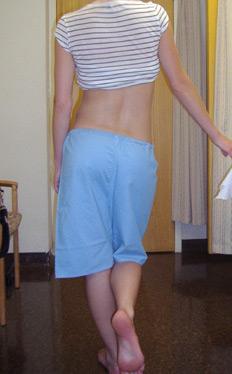
positive apprehension test
Non-specific pain provocation test indicative of an intra-articular problem. The leg is extended over the side of the table in abduction and then externally rotated.
positive modified Thomas' test
The patient sits on the end of the examining bed, and rolls back to a supine position, holding one knee to the chest while the other is hanging supported by the examiner from the end of the bed. The patient holds the knee close to the chest just enough to avoid excessive posterior tilt (lumbar lordosis flattened). The examiner then slowly lowers the free leg. The test is used in order to register whether the hip flexors and especially the iliopsoas are tight in the supine position (positive if the femur is above the horizontal of the table [psoas] and/or the knee extends [rectus femoris and psoas]) and whether stretching of the same muscles is painful and reproduces the known pain.[17]
pain on palpation of inguinal canal
Elicited through the scrotum, preferably in the standing position. The external orifice and the inguinal canal is explored for pain. Any bulging with a Valsalva manoeuvre should also be noted.
pain on palpation of conjoined tendon at pubic tubercle
Palpation of the conjoined tendons (falx inguinalis) of the oblique muscles at the pubic tubercle may elicit pain.
decreased strength and increased pain with hip flexion against resistance (90˚)
Tests strength and functional pain of the iliopsoas with the hip in 90° flexion and the patient in the face-up position.
night pain/rest pain
Atypical pain feature classically seen in patients with infectious or malignant process. [Figure caption and citation for the preceding image starts]: Metastatic lesion of the femoral neck seen on CTFrom the collection of Cedric J. Ortiguera, MD [Citation ends].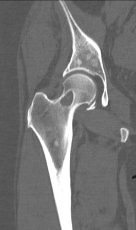 May also signal end-stage osteoarthritis.
Risk factors
strong
previous groin injury
Evidence from football (soccer) and ice hockey suggests that players with a history of groin injury have a 2.5- to 7-fold increased risk of sustaining another groin injury.[10][11] This increased risk is generally attributed to insufficient rehabilitation following the initial injury and/or inherent physiological factors that predispose some individuals to repeat injuries.[11]
higher level of play
Players competing at higher levels may have an increased risk of groin injury due to greater training intensity, higher game demands, and increased training hours.[11]
reduced hip abductor and adductor strength
Lower hip adductor strength, both in absolute terms and relative to hip abductor strength, may lead to decreased muscle capacity and imbalances between the synergistic functions of these muscle groups. This imbalance can increase susceptibility to groin injuries, particularly during movements such as lateral cutting, striding, rapid acceleration or deceleration, and sudden changes in direction.[11]
lower levels of training
Decreased levels of pre-season sport-specific training has been identified as a clear risk factor for groin strain injury in the US National Hockey League.[10] Pre-season training may help in correcting muscle imbalances and enhancing functional recruitment, which helps minimise muscle fatigue. Therefore, athletes with insufficient pre-season training may face a higher risk of injury when training loads increase at the beginning of the season.[11]
weak
increased age
decreased range of motion of the hip
Decreased hip abduction may be a risk factor for groin strain injury, although the evidence is conflicting.[10] One 2017 systematic review indicated strong evidence that total hip rotation below 85° at pre-season screening is a risk factor for developing groin pain. However, it also found that internal rotation, abduction, and extension were not significantly associated with the risk or presence of groin pain.[12]
Use of this content is subject to our disclaimer