Approach
The presentation of groin pain in relation to physical activity can lead to a number of very different diagnoses. Careful attention to the history and examination findings can guide the clinician in determining the correct diagnosis and/or further work-up if indicated. It is important to note that different clinical entities are not mutually exclusive, and they often co-exist.[1]
History
The onset and progression of pain, including whether the pain is acute or chronic, its severity, and any radiation, should be described. Determining the mechanism of injury is helpful, as high-speed movements at long muscle lengths (e.g., cutting, kicking, reaching, jumping) are commonly associated with acute groin injuries. Aggravating and alleviating factors should also be identified to help determine biomechanical or activity-related contributors. A history of trauma, along with details of occupational and recreational activities, should be reviewed. Systemic symptoms, including unexplained weight loss, fatigue, fever, painful urination, and night pain, should be assessed, as they may indicate serious underlying pathology requiring consultant referral.[1]
Physical examination
The physical examination of patients with groin pain should be conducted systematically and interpreted alongside medical history, imaging, and diagnostic injections where appropriate.[1] The precise location of pain (groin, pubis, iliac crest, greater trochanter, thigh, buttock, sacroiliac joint, abdomen, lumbar spine) should be indicated by the patient without the examiner's assistance. Pain arising from multiple sources is common.[1]
Gait assessment
Gait pattern should be assessed to identify abnormalities that may indicate the underlying cause of groin pain.[1] Patterns to assess for include:
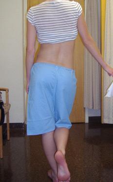
Trendelenburg gait: caused by gluteus medius weakness, leading to pelvic drop on the contralateral side.[Figure caption and citation for the preceding image starts]: Positive Trendelenburg's signFrom the collection of Cedric J. Ortiguera, MD; used with permission [Citation ends].
Coxalgic gait: characterised by rapid unloading of the painful leg during weight-bearing.
Palpation and resistance testing[1]
Palpation and resistance testing should be performed on the adductors, iliopsoas, inguinal canal, and pubic symphysis to identify localised tenderness or pain.
If resistance testing reproduces recognisable pain, and tenderness is present on palpation, extra-articular muscles or tendons are likely involved.
Hip joint evaluation[1]
Passive and active range of motion (ROM) of the hip and knee should be assessed and compared with the contralateral side to identify any pain or limitations.
Hip ROM testing and the FADIR (flexion, adduction, and internal rotation) test help determine intra-articular involvement.
If these tests are pain-free, hip pathology is unlikely.
If pain is present, the hip should be considered as a potential source of groin pain.
Given that hip instability tests have low specificity, rotational and version abnormalities of the femur and tibia should also be assessed, with hip ROM tested in the prone position.
Considering referred pain and serious pathology[1]
In young, active individuals, it is important to consider the possibility of serious pathology, especially if the presentation differs from the key clinical entities for groin pain (adductor, inguinal, iliopsoas, pubic, or hip related).
If pain is suspected to be referred from other areas, examination of the abdomen, lower back, and genitourinary system should be performed at the examiner's discretion.
Imaging
First-line imaging includes plain x-rays (anteroposterior pelvis and anteroposterior/lateral hip). Ultrasound is preferred for suspected inguinal hernias and soft tissue injuries.[1][13]
Second-line imaging includes magnetic resonance imaging (MRI) or MR arthrography (MRA) for assessing femoroacetabular impingement syndrome (FAIS), acetabular dysplasia, femoral torsion, and soft tissue-related groin pain (pubic, iliopsoas, and adductor-related). CT, though useful for 3D hip imaging and FAIS surgical planning, should be used cautiously due to ionising radiation, with low-dose protocols preferred when necessary.[1]
Stress fracture
Superior femoral neck (tension-side) stress fracture and inferior femoral neck (compression-side) stress fracture are serious conditions of the hip that can present with groin pain. Patients may have a history leading to suspicion of overuse injury (e.g., in an endurance athlete or military recruit).
The fulcrum test may help rule out femoral neck stress fractures.[1] Standard x-rays may not reveal fractures. Patients should have an MRI if there is a high clinical suspicion, even if no fracture is visible on plain x-rays.
Groin pain subtypes
The ESSKA-EHPA-ESMA consensus classification of groin injuries in athletes are in principle followed in this topic.[1]
Adductor-related groin pain
History of athletic overuse or traumatic injury.
Presents with pain localised around the insertion of the adductor longus tendon at the pubic bone, which may radiate distally along the medial thigh.
Examination characterised by pain with palpation and pain against resisted adduction of the hip.
Usually a clinical diagnosis, although ultrasound or MRI may be helpful in showing injury to the tendon or the enthesis.
Iliopsoas-related groin pain
History of athletic overuse or traumatic injury.
Presents with pain in the anterior proximal thigh, positioned more laterally compared to adductor-related groin pain.
Examination characterised by pain with palpation of the lower part and the tendon of the iliopsoas, pain on resisted hip flexion test, and/or pain on stretching the hip flexors (modified Thomas test).
Usually a clinical diagnosis; ultrasound or MRI can be helpful showing widening of the tendon.
Inguinal-related groin pain
Caused by a weakening of the posterior inguinal wall.
Presents with pain in the inguinal region that intensifies with activity. In cases of severe pain, discomfort may also occur during coughing, sneezing, or while sitting up in bed.
Pain localised to the inguinal canal region with tenderness upon palpation. No palpable inguinal hernia is noted. Pain is exacerbated during resistance testing of the abdominal muscles or with Valsalva manoeuvre, coughing, or sneezing.
Ultrasound is often useful to demonstrate the soft posterior wall; MRI might be used to rule out other conditions with similar presentations.
Pubic-related groin pain[14]
History of athletic overuse or traumatic injury. Commonly seen in football players.
Presents with pain localised to the symphysis joint and the adjacent bone.
Examination characterised by local tenderness over the pubic symphysis and adjacent bone with palpation. No specific resistance test in addition to palpation reliably provokes symptoms associated with pubic-related groin pain.
MRI may be helpful in identifying pathology.
Hip-related groin pain: FAIS and/or labral tear
Patients may have a family history of hip disease. Labral tear is associated with twisting or pivoting activities, such as ballet, American football, or football (soccer).
Presents with hip, back, buttock, or thigh pain, along with clicking, catching, locking, stiffness, restricted range of motion, or giving way.
Examination is characterised by pain provocation on FADIR (anterior impingement) test, which is sensitive for FAIS and possible labral pathology.
Flexion, abduction, and external rotation (FABER) and straight leg raise tests may also help in identifying intra-articular hip pathology.
Plain x-rays help identify cam or pincer morphology. MRI or MRA is recommended for assessing labral tears, femoral torsion, and prognostic factors for hip preservation surgery.
Hip-related groin pain: acetabular dysplasia and/or hip instability
Presents with anterior, posterior, or lateral/global instability or pain due to acetabular morphology variations. Patients may have a family history of hip disease.
Hip-related groin pain: other conditions without a distinct osseous morphology[15]
Includes isolated or combined involvement of the labrum, cartilage, or ligamentum teres, including various overlapping pathologies such as tears, cysts, erosions, or hypertrophy.
Typically suspected in patients with persistent hip pain despite normal x-rays.
Diagnostic injections
Pain relief obtained from diagnostic intra-articular injections with an anaesthetic and/or corticosteroid can reliably determine the aetiology as being intra-articular (labral tear, chondral lesion) or extra-articular (tendinitis/bursitis, muscular, intra-abdominal, lumbar). The local anaesthetic intra-articular hip injection should be guided by ultrasound or fluoroscopy.[1]
Use of this content is subject to our disclaimer