Images and videos
Images


Age-related macular degeneration
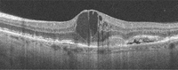
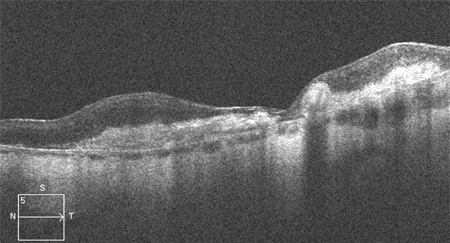
High resolution optical coherence tomography image showing subretinal and intraretinal fluid
Reproduced from Scheie Eye Institute's patient image database; used with permission
See this image in context in the following section/s:

Age-related macular degeneration
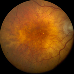
Late AMD with central geographic atrophy (Age-Related Eye Disease Study Group [AREDS] category 4)
Reproduced from Scheie Eye Institute's patient image database; used with permission
See this image in context in the following section/s:
Age-related macular degeneration
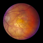
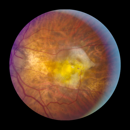
Fibrovascular scar from end-stage AMD (Age-Related Eye Disease Study Group [AREDS] category 4)
Reproduced from Scheie Eye Institute's patient image database; used with permission
See this image in context in the following section/s:

Age-related macular degeneration
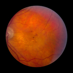
Intermediate AMD (Age-Related Eye Disease Study Group [AREDS] category 3)
Reproduced from Scheie Eye Institute's patient image database; used with permission
See this image in context in the following section/s:

Age-related macular degeneration
Late AMD with choroidal neovascularisation with exudation (Age-Related Eye Disease Study Group [AREDS] category 4)
Reproduced from Scheie Eye Institute's patient image database; used with permission
See this image in context in the following section/s:
Age-related macular degeneration
Optical coherence tomography angiography image showing hyper-reflective lesion which corresponds with a network of choroidal neovascularisation; this has proliferated in between the neurosensory retina and retinal pigment epithelium
From the collection of Sajjad Mahmood MA, MB BCHIR, FRCOphth; used with permission
See this image in context in the following section/s:
Age-related macular degeneration
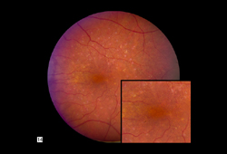
Early AMD (Age-Related Eye Disease Study Group [AREDS] category 2)
Reproduced from Scheie Eye Institute's patient image database; used with permission
See this image in context in the following section/s:
Age-related macular degeneration
High resolution optical coherence tomography image showing hyper-reflective scar
Reproduced from Scheie Eye Institute's patient image database; used with permission
See this image in context in the following section/s:
Age-related macular degeneration
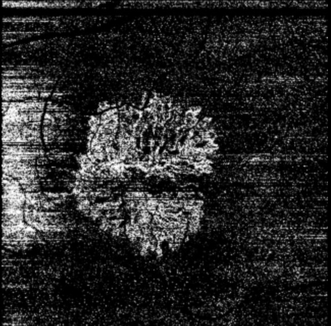
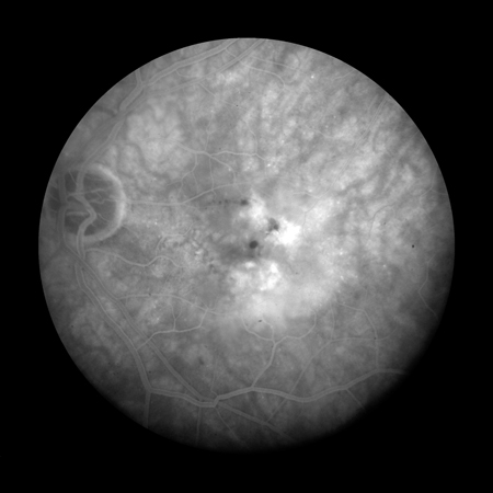
Fluorescein angiogram showing active choroidal neovascularisation
Reproduced from Scheie Eye Institute's patient image database; used with permission
See this image in context in the following section/s:

Age-related macular degeneration
Optical coherence tomography image showing outer retinal hyper-reflective lesion, subretinal and intraretinal fluid
From the collection of Sajjad Mahmood MA, MB BCHIR, FRCOphth; used with permission
See this image in context in the following section/s:
Use of this content is subject to our disclaimer