Approach
It is important to first exclude other vascular diseases, such as atherosclerosis, emboli, and autoimmune diseases, before diagnosing Buerger's disease. Diagnosis is often made after the exclusion of other vascular diseases.
History
Patients are almost invariably smokers and may report episodes of superficial venous thrombophlebitis. Diabetes mellitus, hypertension, and hypercholesterolaemia are notably absent.
The lower limb is often painful and can be eased by hanging the leg over the edge of the bed at night. This suggests ischaemia and is not specific for Buerger's disease. Claudication of the arch of the foot may be described. A history of recurrent superficial thrombophlebitis of either the arms or the legs may be given.
One Japanese survey reported the following symptom frequency in patients with Buerger's disease:[24]
Paraesthesia/cold sensation/cyanosis (37%)
Plantar claudication (15%)
Sural claudication (16%)
Rest pain (10%)
Ulceration/gangrene (19%).
The same study identified involvement of the ulnar artery (12%), anterior tibial artery (41%), and posterior tibial artery (40%) in patients with Buerger's disease.[24]
A history of recurrent episodes of large joint arthritis before their arterial occlusion presentation may be given by 12.5% of patients.[25] Single-joint inflammation is most often described affecting the wrists and knees, lasting up to 2 weeks. The diagnosis of Buerger's disease is often not made until 10 years after the joint symptoms.
Physical examination
Critical ischaemia is defined as gangrene or rest pain lasting >2 weeks and requiring regular opioid analgesia. Its definition may also include ankle pressure <50 mmHg. People with non-critical ischaemia present with either claudication or new onset of rest pain.
An acutely ischaemic limb is a common presentation. The limb is cold with absent infrapopliteal pulses or absent brachial and distal forearm pulses. A sinus heart rhythm is detected. Gangrene or ulceration of the distal phalanges may be noted. [Figure caption and citation for the preceding image starts]: Fingertip ulceration in a woman who smokesFrom the collection of Matthew J. Metcalfe and Alun H. Davies [Citation ends].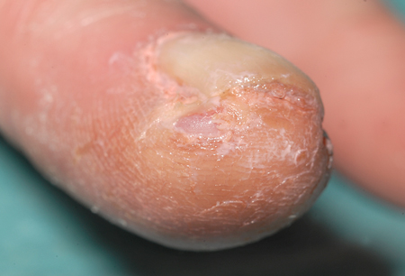 Upper limb involvement is clinically evident in 50% of patients.
The Allen test may detect Buerger's disease in 63% of patients.[5] The Allen test is performed by occluding both radial and ulnar arteries and observing whether the patient's hand becomes ischaemic. The pressure on the radial and ulnar arteries is then released 1 artery at a time. The release of each artery should reperfuse the hand individually. A negative Allen test reveals no ulnar or radial artery occlusion. An abnormal test in a young patient is highly suggestive of Buerger's disease. A positive test in a patient with lower limb disease may indicate the presence of upper limb disease.
Superficial thrombophlebitis is present in 40% to 60% of patients.[26][27]
Laboratory investigations
Investigations should be aimed at ruling out other causes of vascular disease. Blood glucose should be normal, and Buerger's disease can be excluded in patients with diabetes. Biochemical evidence of renal failure (elevated urea and creatinine) may suggest the presence of an autoimmune disease. Erythrocyte sedimentation rate, C-reactive protein, anti-nuclear antibody, rheumatoid factor, anti-neutrophilic cytoplasmic antibody (ANCA), and complement measurements are often normal.[28] However, one study reported 64% incidence of high anti-nuclear antibody levels with positive rheumatoid factor, and 42% incidence of increased anti-nuclear antibody and erythrocyte sedimentation rate levels, in patients with Buerger’s disease.[29] Anti-cardiolipin antibodies may be elevated and are associated with periodontal destruction.[22] Anti-centromere antibodies should be normal to exclude calcinosis, Raynaud's phenomenon, oesophageal dysmotility, sclerodactyly, and telangiectasia (CREST) syndrome. Topoisomerase I antibodies (Scl-70) should be normal to exclude systemic sclerosis (scleroderma). Full blood count may indicate evidence of infection or myeloproliferative disease. Coagulation studies should be normal to exclude a hypercoagulable state. Thrombophilia screen should exclude protein C, protein S, and anti-thrombin III deficiencies.
Arterial Doppler
An arterial Doppler confirms the absence of infrapopliteal, brachial, or distal pulses.
Radiological imaging
Arterial duplex, CT angiography, magnetic resonance angiography, and digital subtraction angiography are all useful radiological imaging techniques that show medium and small vessel occlusion, often with 'corkscrew'-shaped collateral vessels (Martorell's sign) seen.[30] Digital subtraction angiography, CT angiography, and magnetic resonance angiography identify diseased vessels. Arterial duplex identifies non-atherosclerotic occluded vessels. Digital subtraction angiography also demonstrates normal non-atherosclerotic proximal arteries and shows occluded distal small and medium-sized vessels. The choice of imaging technique depends is guided by local availability. CT angiography and magnetic resonance angiography may be better for distal vessel disease.
An echocardiogram should be completed to rule out a cardiac source of distal embolism.
Tissue biopsy
Arterial biopsy may aid diagnosis but should be avoided in ischaemic tissue. In the acute phase, active inflammation of all three vessel layers is seen. In the chronic phase, the thrombus is organised with revascularisation of the medial and adventitial layers. The elastic lamina is preserved.
Use of this content is subject to our disclaimer