Investigations
1st investigations to order
cognitive testing
Test
Cognitive test performance should be recorded at initial assessment and then every 6 months. It is not unusual for cognitive test scores to be in the non-impaired range for up to 1 year after presentation and to decline steadily for about 3-4 years before the patient becomes untestable. These are given by specialists and neuropsychologists, and assist in the diagnosis by documenting a predominance of deficits in the executive and/or language domains of cognition, with relative sparing of memory, orientation, and praxis.
The mini-mental state examination (MMSE) remains the most widely used (and best understood) simple cognitive test. However, patients with frontotemporal dementia (FTD) can score above the cut-off score of 24/30 even if they have significant cognitive impairment, so it is of limited usefulness for detecting FTD. Therefore, other cognitive tests are usually favoured in practice for FTD.
The Montreal Cognitive Assessment (MoCA) can be used to assess for decline in cognition over time; some subsets may give information about poor frontal lobe functioning and track progression.[84][85]
The Addenbrooke’s Cognitive Examination III (ACE III) is a multi-domain test of attention, memory, language, fluency, and visuospatial functions. Maximum total score is 100 with sub-scores for various domains also calculated. It has been validated as a screening tool for cognitive deficits in FTD and Alzheimer’s disease.[86]
The Frontal Assessment Battery (FAB), is a bedside test that can be administered in 10 minutes and is helpful for screening and identifying frontal lobe dysfunction.[87]
The Executive Interview (EXIT) is a useful bedside screening tool for detecting possible frontal lobe dysfunction. It comprises a short screen of 25 items, and takes approximately 15 minutes.[88] The Quick EXIT is an abridged, 14-item version of the original EXIT that was developed by omitting 11 items that were assessed to fit the scale less well.[89]
The Frontal Behavioural Inventory (FBI) is a 24-item, quantifiable questionnaire completed by interviewing an informant.[90] It is aimed at identifying early behavioural and personality changes in behavioural variant FTD (bvFTD).[91]
The Cambridge Behavioural Inventory Revised (CBI-R) is an informant-based questionnaire used to evaluate behavioural symptoms in neurodegenerative diseases, including FTD.[92]
The Sydney Language Battery (SYDBAT) is a test to characterise language deficits in primary progressive aphasia (PPA) variants.[93][94] Four language subtests (naming, word comprehension, repetition, and semantic association) are assessed. A speech language therapist may be involved for a comprehensive assessment of language and devising interventions and compensatory strategies in patients with PPA.[95]
Neurobehavioural scales may also be of use in differentiating from other forms of dementia.[96]
The course of FTD differs from that of most other forms of dementia largely because memory problems are not severe in early stages. Most dementia rating scales are biased towards detection of worsening in Alzheimer's disease. The frontotemporal rating scale (FRS) was developed specifically for FTD, and can detect functional deterioration over 12 months.[97] The degree of dementia severity is more accurately estimated by the FRS than by conventional dementia rating scales.
The Ekman 60 faces test is a test used for checking facial emotion recognition.[98] Poor emotional processing is detectable in patients with FTD and is an important clinical feature in bvFTD and semantic dementia.[99] Emotion recognition can be significantly impaired in bvFTD in comparison to Alzheimer’s disease across all emotions other than happiness.[100] Additionally, recognition of anger is found to be more impaired in FTD than in Alzheimer’s disease, while deficit in recognition of fear is more characteristic of Alzheimer’s disease.[101]
More comprehensive neuropsychological testing of multiple domains is helpful, beyond the initial cognitive tests described earlier in this section, particularly in the early stages when the manifestations of FTD are subtle. Examples of formal test batteries that are appropriate in this setting include the Delis-Kaplan Executive Function System (D-KEFS), Executive and Social Cognition Battery (ESCB), and selected items from the Wechsler Adult Intelligence Scale (WAIS).[102]
Result
disproportionately poor performance in test-taking behaviour and/or in tests of executive functioning; poor emotional processing and impaired facial expression recognition tests; MMSE score often in normal range; in MoCA, frontal subsets may show abnormality
brain MRI
Test
MRI is the key imaging modality for diagnosing FTD and should be ordered if a diagnosis of frontotemporal dementia is suspected.[104][106][Figure caption and citation for the preceding image starts]: Neuroimaging patterns associated with behavioural variant FTD (bvFTD) and nonfluent variant primary progressive aphasia (nfvPPA). Structural MRI and FDG-PET demonstrating the variability in patterns of atrophy and hypometabolism in FTD. In the case of bvFTD, significant bilateral frontal lobe atrophy and hypometabolism is seen. In the case of nfvPPA, atrophy and hypometabolism is lateralised and is greatly impacting the left frontal lobe more so than the right.Peet BT et al. Neurotherapeutics 2021 Apr; 18 (2): 728-52; used with permission [Citation ends].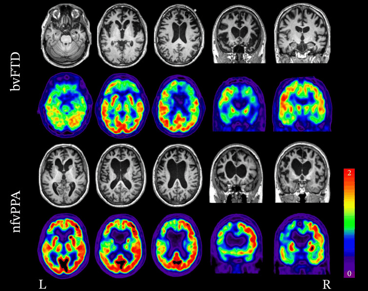 [Figure caption and citation for the preceding image starts]: Coronal T1-weighted MRI (a, b) and fused FDG-PET MRI (c) in a patient with semantic dementiaBhogal P et al. Eur Radiol 23, 3405-17 (2013); used with permission [Citation ends].
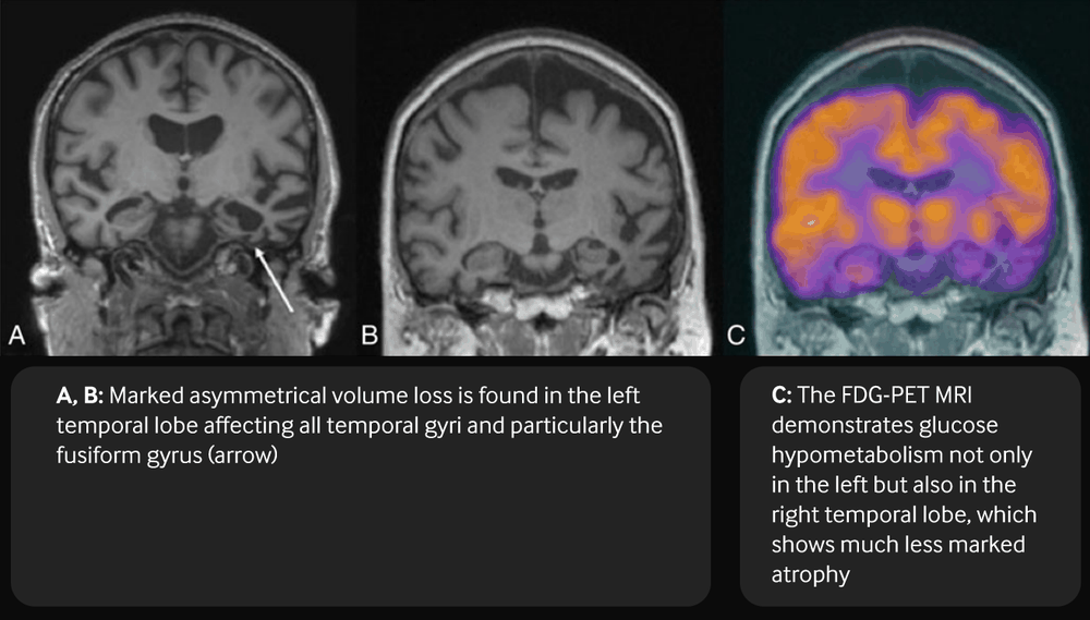
Result
focal atrophy in the frontal and/or anterior temporal lobes; frequently the atrophy is characterised by left-right asymmetry
brain CT
Test
CT should be ordered if a diagnosis of FTD is suspected but MRI is not available or is contraindicated. CT can be used to exclude structural abnormalities such as masses or subdural haematoma that may present with frontal lobe dysfunction, but it may also show atrophy indicative of frontotemporal dementia.[104][106]
Result
focal atrophy in the frontal and/or anterior temporal lobes; frequently the atrophy is characterised by left-right asymmetry
FBC
Test
Ordered to rule out anaemia.
Result
usually normal
serum CRP
Test
Ordered to screen for inflammatory conditions.
Result
usually normal
serum thyroid-stimulating hormone (TSH)
Test
Ordered to rule out hyperthyroidism. TSH is low in hyperthyroidism.
Result
usually normal
free thyroxine (T4)
Test
Ordered to rule out hyperthyroidism. Free T4 is elevated in hyperthyroidism.
Result
usually normal
metabolic panel
Test
Ordered to exclude abnormal sodium, calcium, and glucose levels.
Result
usually normal
serum urea
Test
Ordered to rule out renal failure as a cause of cognitive decline.
Result
usually normal
serum creatinine
Test
Ordered to rule out renal failure as a cause of cognitive decline.
Result
usually normal
LFTs
Test
Ordered to rule out liver failure as a cause of cognitive decline.
Result
usually normal
serum vitamin B12 levels
Test
Ordered to rule out cognitive decline secondary to megaloblastic/pernicious anaemia.
Result
usually normal
serum folate levels
Test
Ordered to rule out cognitive decline due to folate deficiency.
Result
usually normal
syphilis serology
Test
May be positive in syphilis.
Result
usually normal
HIV testing
Test
Positive in dementia in HIV.
Result
usually normal
serum enzyme-linked immunosorbent assay
Test
May be positive for antibodies to Borrelia burgdorferi in Lyme disease.
Result
usually normal
Investigations to consider
brain fluorodeoxyglucose (FDG)-PET/CT
Test
FDG-PET/CT can help differentiate frontotemporal dementia from Alzheimer’s disease and dementia with Lewy bodies.[103][104][105] It is most helpful when combined with MRI.[104][Figure caption and citation for the preceding image starts]: Neuroimaging patterns associated with behavioural variant FTD (bvFTD) and nonfluent variant primary progressive aphasia (nfvPPA). Structural MRI and FDG-PET demonstrating the variability in patterns of atrophy and hypometabolism in FTD. In the case of bvFTD, significant bilateral frontal lobe atrophy and hypometabolism is seen. In the case of nfvPPA, atrophy and hypometabolism is lateralised and is greatly impacting the left frontal lobe more so than the right.Peet BT et al. Neurotherapeutics 2021 Apr; 18 (2): 728-52; used with permission [Citation ends].[Figure caption and citation for the preceding image starts]: Coronal T1-weighted MRI (a, b) and fused FDG-PET MRI (c) in a patient with semantic dementiaBhogal P et al. Eur Radiol 23, 3405-17 (2013); used with permission [Citation ends].
Result
focal hypo-metabolism in the frontal and/or anterior temporal lobes; frequently asymmetrical
brain perfusion single-photon emission computed tomography (SPECT)
Test
SPECT may detect hypoperfusion but is generally considered less helpful than fluorodeoxyglucose (FDG)-PET/CT in the initial imaging of suspected frontotemporal dementia.[104]
Result
focal hypo-perfusion in the frontal and/or anterior temporal lobes; frequently asymmetrical
brain amyloid PET/CT
Test
May be ordered to differentiate frontotemporal dementia from Alzheimer’s disease.[104]
Result
usually normal
brain biopsy
Test
Neuropathological examination is the standard for definitive diagnosis. Specimens may be obtained by brain biopsy, but this is generally not recommended. Therefore, pathological confirmation is typically obtained at the end of life, and is particularly valuable in the characterisation of familial dementia.
Result
on gross examination, regional atrophy predominantly affecting the frontal and/or temporal lobes is found; molecular neuropathology allows most cases of frontotemporal dementia to be placed into one of three molecular subgroups: frontotemporal lobar degeneration with tau (30% to 50% of cases), TAR DNA-binding protein 43 (TDP-43), or fused in sarcoma, Ewing’s sarcoma, and TATA-binding protein-associated factor 15 (FET) protein accumulation
genetic testing
Test
Family history of frontotemporal dementia (FTD) is a good indicator of whether genetic testing is appropriate. Mutations in the GRN and MAPT genes are present almost exclusively in patients with a strong family history, whereas C9orf72 expansion can occur in apparently sporadic disease.[117] Genetic testing is desirable for all patients with probable or possible behavioural variant FTD (bvFTD), and in patients with suspected bvFTD with strong psychiatric features and at least one affected family member. Screening for C9orf72 mutations should take place for all patients with suspected FTD with prominent psychiatric symptoms or family history of late-onset primary psychiatric disorder (even if full diagnostic criteria are not met).[28] Genetic testing (for MAPT, GRN, and C9orf72) should also be performed if the results could contribute to decisions about pregnancy.
Genetic testing should be considered within the context of a clinical genetic service with the capacity to provide educational and psychological support for affected families. Pre- and post-test counselling is recommended. Genetic counselling assesses family history, and advises on genetic testing and its impact. It helps individuals and families understand the potential risks and implications of FTD, particularly familial types, and facilitates decisions about genetic testing and management of the condition. Following testing, counselling helps with interpreting results, and provides support and information about inheritance patterns and potential risks for other family members.[118][119]
Result
demonstration of mutations in the MAPT, GRN, or C9orf72 genes typically; mutations in CHMP2B and other genes are rare; analysis may identify a gene pertinent to a different dementia (such as Alzheimer's disease), in which circumstance the case is an FTD phenocopy
connective tissue panel
Test
Ordered if indicated; positive results may rule out frontotemporal dementia.
Result
usually normal
serum erythrocyte sedimentation rate
Test
Ordered to screen for inflammatory conditions.
Result
usually normal
electroencephalogram (EEG)
Test
EEG use in dementia is endorsed when there has been a history of seizures or altered consciousness, raising the possibility of epilepsy, encephalopathy, or atypical subacute course.[106][120] Patterns of lateralised or bilateral epileptiform abnormalities, or slow (sometimes triphasic and periodic) waves support a diagnosis of late-onset epilepsy, prion disease, or toxic, metabolic, septic, autoimmune, and anoxic encephalopathies.[106][121][122][123]
Result
usually normal
electromyogram (EMG)
Test
An EMG may be indicated if symptoms and signs suggestive of amyotrophic lateral sclerosis are noted. In lower motor neuron dysfunction, an EMG may show evidence of chronic neurogenic change (large motor unit potentials of increased duration and/or increased amplitude), and evidence of ongoing denervation (fibrillation potentials or positive sharp waves, or fasciculation potentials).[126]
Result
usually normal
Emerging tests
fluid biomarkers
Test
There is growing interest in exploring potential fluid biomarkers in cerebrospinal fluid (CSF), serum and plasma for diagnostic, prognostic, and staging purposes.
There is no specific single fluid biomarker that uniquely identifies frontotemporal (FTD) pathologies associated with sporadic cases. Various markers have been and continue to be studied. Using a combination of markers may potentially increase diagnostic accuracy.[129]
Compared with Alzheimer’s disease, FTD is characterised by higher levels of CSF Aß42, and lower t-tau/Aß42 and p-tau/Aß42 values. The ratio of p-tau/Aß42 appears to be the most sensitive and specific biomarker for discriminating FTD with primary language disturbances from Alzheimer’s disease.[130] It has also been shown that plasma p-tau-181 and p-tau-217 are raised in Alzheimer’s disease but not FTD, except for certain MAPT mutations.[131][132] In patients with TAR DNA-binding protein 43 (TDP-43) proteinopathy, there is a reduction in the ratio of p-tau181 to total tau.[133] Multiple other fluid biomarkers are being researched and may become clinically available in the future.
Result
CSF Aß42 elevated compared with Alzheimer’s disease, t-tau/Aß42 and p-tau/Aß42 values lower than in Alzheimer’s disease, plasma p-tau-181 and p-tau-217 usually normal, ratio of p-tau181 to total tau reduced in patients with TDP-43 proteinopathy
Use of this content is subject to our disclaimer