Images and videos
Images
Iron deficiency anemia
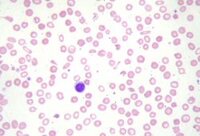
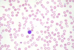
Peripheral blood smear demonstrating some changes often seen with iron deficiency anemia. Note that many of the red cells are microcytic (compare size of red cell with the lymphocyte nucleus) and hypochromic (wide central pallor). There are some pencil forms
From personal collection of Dr Rebecca Fischer Connor; used with permission
See this image in context in the following section/s:
Iron deficiency anemia
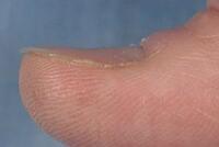
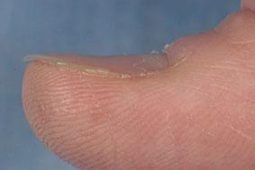
Koilonychia
Reproduced with permission from Bickle Ian. Clinical exam skills: Hand signs BMJ 2004;329:0411402
See this image in context in the following section/s:
Videos
 Venepuncture and phlebotomy: animated demonstration
How to take a venous blood sample from the antecubital fossa using a vacuum needle.
Use of this content is subject to our disclaimer