History and exam
Key diagnostic factors
common
presence of risk factors
Key risk factors are trauma to the skin, outdoor occupation or hobbies, animal bites or scratches, alcoholism, diabetes mellitus, HIV, or COPD.
primary skin lesion
A nodular non-tender lesion develops 1 to 12 weeks after traumatic fungal inoculation. This lesion usually ulcerates. Patients with fixed cutaneous sporotrichosis may develop verrucous-like lesions that may not ulcerate. Skin lesions are non-tender and pain is extremely uncommon.[Figure caption and citation for the preceding image starts]: Ulcerated primary sporotrichosis lesionFrom the collection of Richard J. Hamill, MD, Baylor College of Medicine, Houston, TX [Citation ends].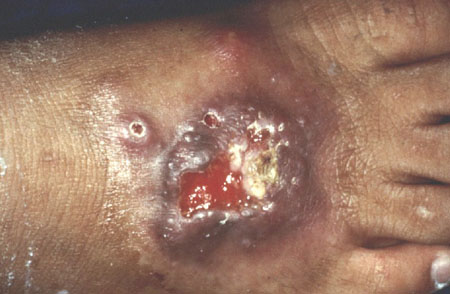
nodular lymphangitis lesions
A 'sporotrichoid' characteristic spread of the infection across proximal lymphatic channels is common. Nodular non-tender lesions, similar to the primary skin lesion, develop along the lymphatics that may also ulcerate.[Figure caption and citation for the preceding image starts]: Ascending 'sporotrichoid' distribution of skin lesions across the proximal lymphatic channelsFrom the collection of Richard J. Hamill, MD and Edward Septimus, MD, Baylor College of Medicine, Houston, TX [Citation ends]. [Figure caption and citation for the preceding image starts]: Proximal lymphangitic spread of primary lesionFrom the collection of Richard J. Hamill, MD, Baylor College of Medicine, Houston, TX [Citation ends].
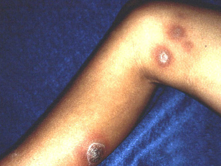
Other diagnostic factors
common
respiratory symptoms
joint swelling/erythema
Joint swelling, erythema, and associated joint effusion occur in osteoarticular sporotrichosis. Monoarthritis, oligoarthritis, or polyarthritis may occur.
The knee is the joint most frequently affected, followed by the wrist, hand, elbow, and ankle. There is sparing of the shoulder, hip, and spine. Associated bursitis, tenosynovitis, and nerve entrapment syndromes such as carpal tunnel syndrome may develop.
male 30 to 60 years old
Typical patient demographics for pulmonary sporotrichosis. In non-pulmonary forms of sporotrichosis, men or women are more commonly affected depending on the geographic region.
uncommon
fever
May occur in patients with pulmonary and disseminated sporotrichosis. Not present in lymphocutaneous and osteoarticular forms of sporotrichosis.
lymphadenopathy
Regional lymphadenopathy may rarely occur.
meningitis-like symptoms
Meningeal sporotrichosis follows a chronic indolent clinical course, similar to that seen with meningeal histoplasmosis and coccidioidomycosis.[1]
Risk factors
strong
trauma to the skin
Traumatic inoculation of the fungus to the skin causes lymphocutaneous/cutaneous sporotrichosis. Patients do not always recall sustaining trauma to the affected area (recollection of trauma in 10% to 62% of patients in different reported series).[11][26] Hence, because microscopic skin abrasions that a patient may not notice or remember are sufficient to introduce the fungus and cause infection, lack of major trauma or penetrating injury should not lower the clinical suspicion for sporotrichosis.
outdoor occupation/hobbies
Sporothrix schenckii is abundant in soil, wood, sphagnum moss, thorns, decaying vegetation, and hay. Hence, people who engage in hobbies or occupations that involve the outdoors such as gardening, landscaping, topiary production, Christmas tree farming, and hay baling are at risk for developing sporotrichosis.
animal bites and scratches
Bites and scratches from various animals have been reported to cause sporotrichosis. Cats and armadillos are the most common animals implicated in zoonotic transmission of sporotrichosis to humans. Cats may develop severe, often fatal, sporotrichosis infections with ulcerative skin lesions that contain large numbers of fungal organisms, which can be directly introduced to humans following cat scratches. On the other hand, armadillos do not develop Sporothrix infections but may carry the fungus, and by inflicting scratches on armadillo hunters, they may inoculate the fungus into the skin. Insect, dog, and rodent bites have also been implicated in zoonotic sporotrichosis transmission.
alcohol use disorder
There is a strong association of excessive alcohol use with development of pulmonary, osteoarticular, and disseminated sporotrichosis. The underlying mechanism accounting for this predilection is unknown, but may be related to an altered T-helper 1 cell response.[27]
diabetes mellitus
Diabetes mellitus is associated with extracutaneous forms of sporotrichosis. It is unclear what the underlying mechanism for this susceptibility is, but it may relate to the functional neutrophil impairment that occurs during hyperglycaemia.
HIV/AIDS
Patients with HIV develop cutaneous/lymphocutaneous sporotrichosis following traumatic inoculation to the skin as normal hosts do, although in HIV-infected individuals, skin lesions are usually more extensive. In addition, patients with HIV can develop isolated osteoarticular, pulmonary, and meningeal sporotrichosis as well as disseminated cutaneous and visceral sporotrichosis. Most cases of extracutaneous and disseminated sporotrichosis in patients with HIV have been reported with CD4 counts of <100 cells/mm³, whereas patients with HIV and with lymphocutaneous or fixed cutaneous sporotrichosis have mean CD4 counts of >200 cells/mm³.[28] It has been suggested that disseminated sporotrichosis in patients with HIV should be classified as an AIDS-defining illness.[28]
COPD
Risk factor for pulmonary sporotrichosis.
weak
corticosteroid treatment
Risk factor for disseminated sporotrichosis, likely caused by the impairment of neutrophil and T-cell-mediated immune responses in the setting of iatrogenic hypercortisolism.
chemotherapy in patients with haematological malignancy
Risk factor for disseminated sporotrichosis.
haematopoietic stem cell/solid organ transplantation
Risk factor for disseminated sporotrichosis.
tumour necrosis factor (TNF)-alpha inhibitor therapy
A single case of disseminated sporotrichosis has been reported in a patient who received both TNF-alpha antagonists etanercept and infliximab.[29]
Use of this content is subject to our disclaimer