Approach
Clinical assessment is focused on a history of hip pain, limp, and a finding of external rotation of the hip on examination.[27] Assessment of weight-bearing ability is vital as this helps to determine the prognosis for avascular necrosis as well as the urgency of surgical intervention. Bilateral hip radiology in both antero-posterior and lateral viewpoints should be requested to confirm diagnosis. [Figure caption and citation for the preceding image starts]: Unstable SCFE of the right hip. Antero-posterior preoperative x-rayImage courtesy of John M. Flynn, MD [Citation ends]. [Figure caption and citation for the preceding image starts]: Unstable SCFE of the right hip. Frog-leg lateral preoperative x-rayImage courtesy of John M. Flynn, MD [Citation ends].
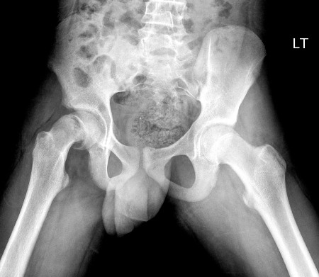
History
Typical presenting features include medial knee, hip, groin, and/or thigh pain. Pain may be referred to these regions. Onset may have been acute or insidious. A history of trauma from falls or a sport injury may be elicited. Remember: with unexplained unilateral knee pain in an adolescent, always consider the hip.
A diagnosis of SCFE has implications for immediate treatment to prevent slip progression and avoid complications such as avascular necrosis or chondrolysis.[1][28] Furthermore, in unstable SCFE circulation may be compromised if any of the retinacular vessels are disrupted. This may require emergency surgical treatment.[29]
Valgus SCFE, defined as posterolateral slippage of the proximal femoral epiphysis on the metaphysis, is more likely to be present in younger and female patients with a higher neck shaft angle than classic posteromedial SCFE cases.[3]
Physical examination
In patients <10 years of age, features of panhypopituitarism, the presence of growth hormone deficiency after supplementation has begun, renal osteodystrophy, and especially hypothyroidism should be sought. Weight should be measured: if <50th percentile, an endocrine disorder should be suspected as a contributory risk factor. If >90th percentile, the child has a significant risk factor for SCFE.
Obligatory external rotation is present when the hip joint is flexed. Passive and active flexion of the hip should be conducted. The affected hip tends to go into external rotation during flexion, due to the resultant anatomical deformity of the proximal femur. Range of movement is typically restricted. The patient may limp and gait is characteristic for the patient to walk with their leg externally rotated. Observational gait analysis is therefore recommended. Trendelenburg's test is positive. The test is performed by the child standing on the affected leg with the knee flexed and the hip extended.[30] The trunk typically leans towards the affected side.
Investigations
Blood tests including metabolic panel, thyroid function tests, and pituitary hormones (including growth hormone) should be obtained if there are features suggestive of an underlying disorder.
Plain x-rays are generally sufficient for diagnosis of SCFE.[31] Bilateral films should be obtained because SCFE often occurs in both hips (up to 60% of cases).[32] Both antero-posterior (AP) and lateral views should be requested. The frog-leg lateral view is often a better diagnostic tool and can show the posteriorly displaced capital femoral epiphysis.[32] Frog-leg lateral radiographs show the Klein line not intersecting the femoral head. Other radiographical findings may include widening of the physis on the affected side, loss of the overlap of the metaphysis of the proximal femur (Capener’s sign), or increased sclerosis of the proximal metaphysis on the AP view, where the slipped epiphysis overlies the metaphysis as a double shadow (blanch sign of Steel).[33][34]
Valgus SCFE is often difficult to recognise on AP films. For this reason, it is important to obtain lateral and contralateral comparison views in children with hip pain.[3][Figure caption and citation for the preceding image starts]: Unstable SCFE of the right hip. Antero-posterior preoperative x-rayImage courtesy of John M. Flynn, MD [Citation ends].[Figure caption and citation for the preceding image starts]: Unstable SCFE of the right hip. Frog-leg lateral preoperative x-rayImage courtesy of John M. Flynn, MD [Citation ends].[Figure caption and citation for the preceding image starts]: Klein lines are drawn along the superior cortex of the femoral neck. A normal Klein line will intersect the epiphysis. An abnormal Klein line does not intersect the epiphysis, as the femoral neck has moved proximally and anteriorly relative to the epiphysisImage courtesy of John M. Flynn, MD [Citation ends].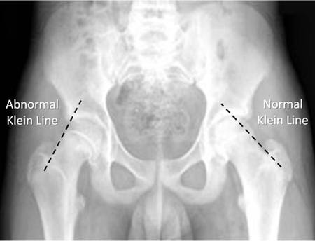
Urgent referral to orthopaedic surgeon
SCFE should be treated surgically as soon as it is recognised. The patient should be advised not to bear weight on the affected hip and should be immediately referred to an orthopaedic surgeon. Early surgical intervention can prevent slip progression and complications such as osteonecrosis.
Use of this content is subject to our disclaimer