Images and videos
Images
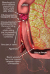


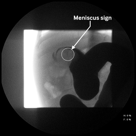
Intussusception
Site of intussusception as revealed by abdominal x-ray, showing the meniscus
From the collection of Dr David J. Hackam; used with permission
See this image in context in the following section/s:

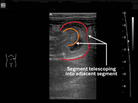
Intussusception
Ultrasound image showing invagination of a segment of bowel into the adjacent segment
BMJ Case Reports. 2009; doi:10.1136/bcr.04.2009.1730; used with permission
See this image in context in the following section/s:

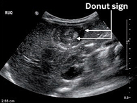
Intussusception
Transverse sonogram of the abdomen showing the donut sign (concentric rings within the lumen of a distended loop of bowel)
Adapted from the Student BMJ. 2008;16:76. Copyright 2010 by the BMJ Publishing Group; used with permission
See this image in context in the following section/s:
Intussusception
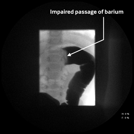
Abdominal x-ray showing impaired passage of barium at site of obstruction due to intussusception
From the collection of Dr David J. Hackam; used with permission
See this image in context in the following section/s:
Intussusception
We are sorry, but we have encountered a problem
Refresh the page and if the problem persists, please contact us.
Contact usBack to BMJ Best Practice