Images and videos
Images
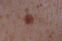

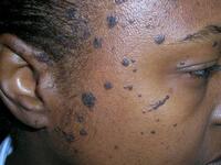

Seborrheic keratosis
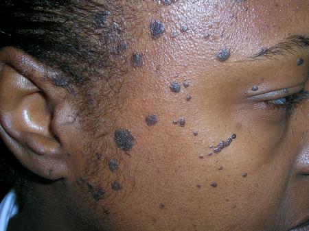
Dermatosis papulosa nigra, a common subtype of seborrhoeic keratosis which is more prevalent in people with brown or black skin (Fitzpatrick skin type IV, V or VI)
Richard Usatine MD/Science Photo Library; used with permission
See this image in context in the following section/s:

Seborrheic keratosis
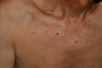
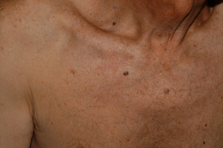
Seborrheic keratosis on the chest of an elderly woman
From the collection of Dr Braun and Dr Kolm, used with permission
See this image in context in the following section/s:
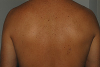
Seborrheic keratosis
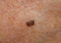
Clinical close-up image of seborrheic keratosis on the back of a 40-year-old man
From the collection of Dr Braun and Dr Kolm, used with permission
See this image in context in the following section/s:

Seborrheic keratosis
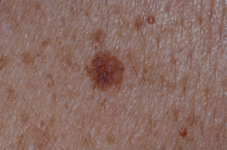
Dermoscopic image of seborrheic keratosis on the chest
From the collection of Dr Braun and Dr Kolm, used with permission
See this image in context in the following section/s:

Seborrheic keratosis
Seborrheic keratosis of the chest: clinical close-up image
From the collection of Dr Braun and Dr Kolm, used with permission
See this image in context in the following section/s:
Seborrheic keratosis
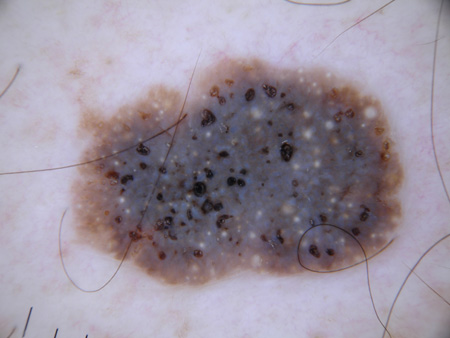
Example of a dark-brown pigmented seborrheic keratosis. Dermoscopic image: see yellowish horn pearls and dark brown holes corresponding to so-called "pseudo-follicular openings"
From the collection of Dr Braun and Dr Kolm, used with permission
See this image in context in the following section/s:
Seborrheic keratosis
Seborrheic keratosis of the chest: clinical overview image
From the collection of Dr Braun and Dr Kolm, used with permission
See this image in context in the following section/s:
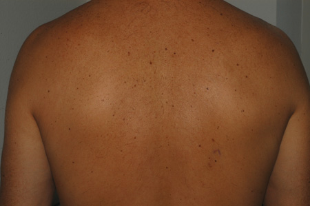
Seborrheic keratosis
Clinical overview image of seborrheic keratosis on the back of a 40-year-old man
From the collection of Dr Braun and Dr Kolm, used with permission
See this image in context in the following section/s:
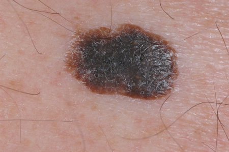
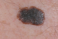
Seborrheic keratosis
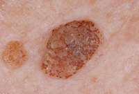
Clinical image of an example of a dark-brown pigmented seborrheic keratosis
From the collection of Dr Braun and Dr Kolm, used with permission
See this image in context in the following section/s:
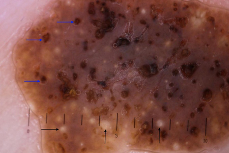
Seborrheic keratosis
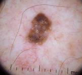
Typical dermoscopic appearance of seborrheic keratosis with milia-like cysts (black arrows) and comedo-like openings (blue arrows)
Primary Care Dermatology Society (PCDS); used with permission
See this image in context in the following section/s:
Use of this content is subject to our disclaimer