Aetiology
The anatomical relationships within the thoracic outlet provide potential areas of neurovascular compression. The areas that typically impart neurovascular compromise include the interscalene triangle, the costoclavicular space, and the subcoracoid space (also called the pectoralis minor space). Congenital, developmental, and traumatic factors may all contribute to anatomical narrowing within the thoracic outlet.
Congenital factors
Congenital factors which may cause thoracic outlet syndrome (TOS) include supernumerary cervical ribs (complete or partial), anomalous (hypoplastic) first ribs, extra-numerary muscles (such as a scalene minimus), and fibrofascial or ligamentous bands (e.g., 'Roos' bands).[22][57][58][59][60][61][62] Any of these elements can result in reduced space within the thoracic outlet, predisposing to symptomatic compression of neurovascular structures.
Developmental factors
Developmental factors relate to changes in the structure of the thoracic outlet which occur over time, as a consequence of growth and daily activity.
Factors such as posture, exercise, work habits, and hobbies influence these changes. For example, weightlifting can cause muscle hypertrophy, while prolonged use of computers and mobile devices may lead to poor posture, characterised by a rounded neck and shoulders. Athletes or workers performing repetitive overhead tasks may experience repeated stretching, distracting, and compressing of neurovascular structures across the thoracic outlet and subcoracoid spaces.[4]
Activities repeated daily for prolonged periods of time may eventually alter musculoskeletal growth, which in turn may alter the relative positions of the first rib, clavicle, scalene muscles, and neurovascular structures. This alteration may decrease the normal aperture of the thoracic outlet and increase compression, leading to symptoms such as pain, paraesthesia, and weakness in nerves; congestion, swelling, discoloration, and thrombosis in veins; and stenosis, aneurysm formation, occlusion, thrombosis, embolism, pain, paraesthesia, loss of pulses, coldness, and weakness in arteries.
Traumatic factors
Hyperextension injuries resulting from rapid acceleration or deceleration of the neck or arm (e.g., whiplash-like injury or falls on the outstretched arm) are well known to be contributing factors in neurogenic TOS. These types of injury may result in microscopic alterations in scalene and pectoralis minor muscles, with fibrosis and varying degrees of chronic muscle spasm. These factors may lead to compression and irritation of the brachial plexus in the interscalene triangle, costoclavicular space, and/or subcoracoid space.[11][12][63][64][65]
Fractures of the clavicle or first rib may result in abnormal bone alignment or callus formation.[7][11][65] The repair of clavicular fractures with plates and screws may result in iatrogenic compression or injury of neurovascular structures at the thoracic outlet.[66][67][68]
Pathophysiology
Knowledge of thoracic outlet anatomy is fundamental to understanding the pathophysiology of thoracic outlet syndrome (TOS).[22][57][58][59][60][61][62][69][70][71][72][73]
Normal thoracic outlet anatomy
The surface landmarks of the thoracic outlet include the clavicle and sternoclavicular joint, the lateral aspect of the sternocleidomastoid muscle, the upper border of the trapezius muscle, the pectoralis major muscle of the upper anterior chest, and the deltoid muscle of the shoulder.
[Figure caption and citation for the preceding image starts]: Surface landmarks of the thoracic outletFrom the collection of Dr. Robert W. Thompson; used with permission [Citation ends].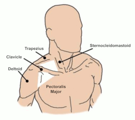
In the supraclavicular space the structures of the thoracic outlet are deep to the scalene fat pad, which contains an abundance of adipose and lymphatic tissues. At a deeper level, the first rib arises from the transverse process of the T1 vertebra and it arcs forward to join the sternal manubrium just inferior and deep to the sternoclavicular joint. The anterior and middle scalene muscles arise from the transverse process of the cervical spine (C3-C6) and pass vertically to attach to the top of the first rib, forming the scalene triangle. The subclavius muscle arises along the lower border of the mid-clavicle with an attachment to the anteromedial first rib as the thickened costoclavicular ligament. The pectoralis minor muscle arises from the anterior costal cartilage of ribs 3-5, passing transversely to attach to the coracoid process of the scapula, immediately above the insertion of the short head of the biceps muscle.
[Figure caption and citation for the preceding image starts]: The subclavian vein and artery pass over the first rib and under the clavicle. The brachial plexus traverses the top of the bony circle to join the artery. The apex of the pleura (cupula) is shown on the left sideReprinted with permission from Elsevier [Citation ends].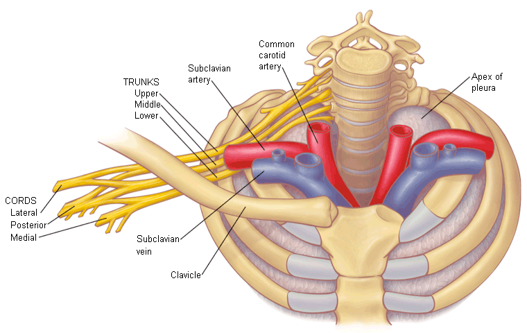
The three spaces of the thoracic outlet are defined by these musculoskeletal structures and the neurovascular components that pass through them:
The supraclavicular scalene triangle, bound by the anterior and middle scalene muscle and first rib
The costoclavicular space, bound by the clavicle, subclavius muscle, anterior scalene muscle, and first rib
The infraclavicular subcoracoid (pectoralis minor) space, bound by the coracoid, pectoralis minor muscle and tendon, and the underlying chest wall
[Figure caption and citation for the preceding image starts]: The scalene muscle triangle is the first major level of neurovascular compressionReprinted with permission from Elsevier [Citation ends].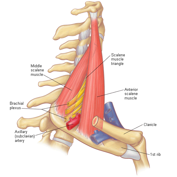
[Figure caption and citation for the preceding image starts]: Lateral view of the neurovascular structures traversing the thoracic outlet, with the clavicle above and the first rib below. The costoclavicular space is the second major site of neurovascular compressionReprinted with permission from Elsevier [Citation ends].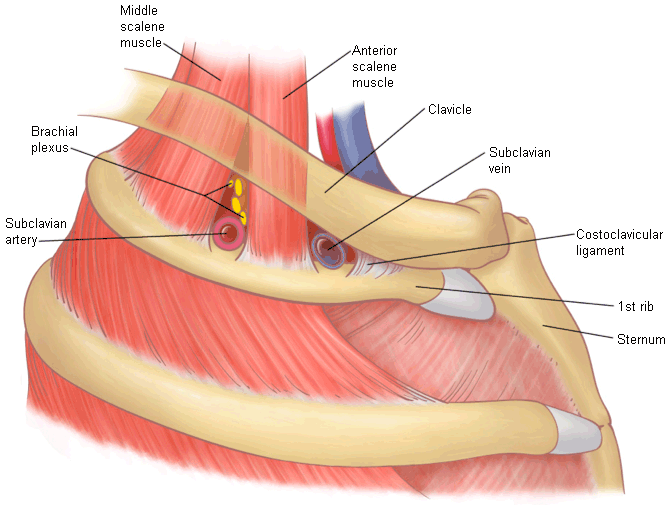
[Figure caption and citation for the preceding image starts]: The neurovascular structures pass behind the pectoralis minor muscle, a third major area of neurovascular compression. The pectoralis minor muscle is a shoulder protractor, which can overcome the rhomboids. The shoulder retracts and alters the thoracic outlet, contributing to muscle imbalance and compression of the brachial plexusReprinted with permission from Elsevier [Citation ends].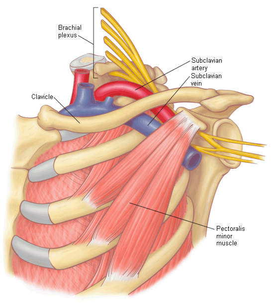
The brachial plexus arises from cervical nerve roots C5-T1 to form three trunks: upper trunk (C5 and C6), middle trunk (C7), and lower trunk (C8 and T1). These nerve roots and trunks pass through the scalene triangle and over the first rib, then branch and fuse further to form the brachial plexus divisions and cords as they pass into the axilla and subcoracoid space. Beyond this level arise the principal peripheral nerves of the upper extremity: median, ulnar, musculocutaneous, axillary, and radial. Other nerves relevant to the thoracic outlet include the phrenic nerve, which passes inferiorly along the fascia of the anterior scalene muscle and under the subclavian vein to the mediastinum, to innervate the ipsilateral hemi-diaphragm; the long thoracic nerve and dorsal scapular nerves, which pass through the middle scalene muscle and posteriorly to innervate the serratus anterior and rhomboid muscles, respectively; and the suprascapular nerve, which arises from the upper trunk of the brachial plexus to pass posteriorly to the scapular notch and then to innervate the supraspinatus and infraspinatus muscles.
The subclavian artery leaves the chest by curving up over the first rib behind the anterior scalene muscle and in front of the brachial plexus and middle scalene muscle. It then passes over the first rib and under the clavicle to enter the axilla beneath the pectoralis minor muscle. Branches of the proximal subclavian artery include the vertebral artery, internal thoracic (mammary) artery, and thyrocervical trunk. The axillary artery gives origin to additional branch vessels in the subcoracoid space (superior thoracic, thoraco-acromial, lateral thoracic, subscapular, and anterior circumflex humeral and posterior circumflex humeral arteries), then continues as the brachial artery to the arm.
The axillary vein arises in the subcoracoid space by joining the basilic, brachial, and cephalic veins of the arm, then passes centrally underneath the clavicle and subclavius muscle where it becomes the subclavian vein. The subclavian vein passes over the first rib through the costoclavicular space in front of the anterior scalene muscle. It turns inferiorly to join the internal jugular vein in forming the brachiocephalic (innominate) vein, which passes behind the head of the clavicle and sternal manubrium into the superior mediastinum.
Anatomical thoracic outlet variations
There are a large number of variations in the musculoskeletal anatomy within the thoracic outlet.[22][57][58][59][60][61][62] The most obvious bony anomalies are supernumerary cervical ribs, which arise from the transverse process of the C7 vertebrae and have embryonic origin within the plane of the middle scalene muscle. Complete cervical ribs have a distal bony attachment to the anterolateral first rib, often forming a joint-like pseudoarthrosis, whereas partial cervical ribs typically terminate in a ligamentous band. Under-developed (hypoplastic) first ribs can appear radiographically similar to cervical ribs but arise from the T1 vertebrae, with either attachment to the lateral second rib or termination in a fibrous band. There are also rare bony variations with bridge-like fusions between the first and second ribs.
Muscle variations include fibres of the posterior part of the lower anterior scalene muscle that can pass over and around the subclavian artery to attach to the extrapleural fascia rather than the first rib, forming a firm sling that can fixate and elevate the artery with muscle contraction. Another prominent muscle variation is the scalene minimus muscle, which originates in the plane of the anterior scalene muscle, passes between various trunks of the brachial plexus, and terminates within the plane of the middle scalene muscle or onto the extrapleural fascia. Sibson’s fascia refers to the thickened extrapleural fascia at the apex of the pleura, which attaches to the internal border of the first rib and the transverse process of C7 and can have varying degrees of thickness in the region of the thoracic outlet, particularly near the C8 and T1 nerve roots. Other musculofascial bands have been described and classified, producing varying degrees of potential neurovascular compression. In the axilla, there can be some degree of variation in the muscular attachment of the pectoralis minor muscle to the coracoid process.[74][75] Another variation is a firm fascial band arising from the latissimus dorsi muscle, crossing over the adjacent neurovascular structures to attach to the pectoralis major, coracobrachialis, or biceps muscles, known as Langer’s axillary arch.[76]
It is crucial to recognise that the anatomy of the thoracic outlet is not static: muscle hypertrophy and post-traumatic changes may influence any potential neurovascular compression. There are also potentially significant changes in the degree of neurovascular compression that may occur with different positions of the neck, shoulder girdle, and upper extremity, as well as with various upper extremity activities. The dynamic nature of thoracic outlet anatomy is thereby viewed as a particularly important feature of TOS.
Neurogenic TOS
Repetitive stress injuries and hyperextension injuries of the neck have been found to cause acute microtearing, inflammation, and persistent spasm of the scalene muscles. Subsequently, fibrosis and tightness of the scalene muscles develops, which also creates narrowing of the interscalene triangle and compression of the neurovascular bundle. These changes may lead to a cycle of sustained muscle spasm resulting in pain, which leads to a feedback mechanism where further spasm results following the painful stimulus. Anatomical abnormalities also serve as a critical predisposing factor, enhancing the likelihood of neurovascular compression when combined with muscle injuries.[1] Ultimately this leads to both hypertrophy of the scalene muscles as well as microscopic skeletal muscle changes, with conversion to a large disproportion of slow-sustained contraction muscle-type fibres.[77][78] The presence of congenital or acquired structural factors further promotes nerve compression. The net result is increased compression of neural structures resulting in the symptoms of upper extremity pain, paraesthesia, and weakness.
Venous TOS
Venous TOS results from progressive compression of the subclavian vein as it crosses the costoclavicular space. The compression can be multifactorial caused by the clavicle, first rib, the anterior scalene muscle, and the subclavius muscle/costoclavicular ligament.
The most common pathological mechanism relates to forceful repetitive activity with the arm elevated or overhead, resulting in hypertrophy of the anterior scalene and subclavius muscles. As these muscles enlarge the space between them may be reduced. The normal function of these muscles is to draw the clavicle and first rib into proximity. As the muscles enlarge, they can further narrow the space between the clavicle and first rib. Hypertrophy has also been described in the pectoralis minor muscle, which can contribute to positional venous obstruction in the subcoracoid space.[17][79]
The stresses of tension developed by the scalene and subclavius muscles may lead to changes in the periosteal growth of the medial first rib, resulting in osteophytic changes, exostoses, and radiographically occult fractures. Soft tissue changes around the altered first rib can in turn further reduce the costoclavicular space.[80]
Once the compression results in a critical impingement on the subclavian vein, progressive fibrous stenosis occurs with repetitive vein injury and as the scar tissue constricts the vein further. During this time venous flow from the arm is preserved by concurrent expansion of collateral vein pathways. Once the blood flow in the vein becomes reduced to the point where blood may coagulate and obstruct the vein entirely, the thrombus may propagate distally into the axillary vein where collateral vein origins may also become occluded. This often results in dramatic spontaneous upper extremity congestion, swelling, discoloration, and pain, extending from the shoulder to the hand. This condition has been termed 'effort thrombosis' of the subclavian vein or the Paget-Schroetter syndrome.[79]
Some patients will have intermittent positional compression of the axillary or subclavian veins that is sufficient to cause symptoms of upper extremity venous congestion in the absence of venous thrombosis. This has been termed McCleery’s syndrome.[15][16]
Venous TOS may complicate creation and maintenance of haemodialysis access, potentially related to high venous flow and previous subclavian vein injury from catheter placement. Arm swelling, diminished arteriovenous fistula function, or repeated vascular access thrombosis may reflect subclavian vein compression at the costoclavicular junction.[81][82][83][84][85][86][87]
Arterial TOS
The same congenital factors and muscle alterations that contribute to neurogenic TOS also occur with arterial TOS, as the subclavian artery courses through the thoracic outlet adjacent to the brachial plexus.
There is a very strong correlation between the development of arterial TOS and the presence of bony abnormalities, particularly cervical ribs (present in 75% of patients).[1][18][55]
Central to the definition of arterial TOS is chronic vascular injury with damage to the subclavian artery. This type of injury is from chronic external compression at the thoracic outlet and the ensuing haemodynamic changes. The arterial wall damage may take the form of a fixed stenosis (chronic narrowing) or development of aneurysmal enlargement of the artery. This type of aneurysmal enlargement typically occurs within 2-3 cm of the point of chronic stenosis, due to the magnified haemodynamic forces and flow turbulence in this region, and is thereby referred to as post-stenotic dilatation.[54]
The presence of these lesions allows for thrombus to form along the arterial wall within the damaged dilated segment of the subclavian artery. Because of the force of arterial blood flow, mural thrombus may dislodge and be carried in the bloodstream to result in blockage of downstream arteries in the arm or hand. Occasionally, the subclavian artery may become obstructed by thrombus with the entire arm becoming ischaemic. In some instances, thrombus may extend retrograde toward the more central subclavian and vertebral arteries, with embolisation to the brain resulting in a vertebrobasilar (posterior brain) stroke.[23][24][25][88]
Intermittent compression of the axillary artery can occur in the subcoracoid space, opposite the level of the head of the humerus. Repetitive compression at this site is typically seen in high-performance overhead athletes (e.g., volleyball, baseball, softball), and can lead to similar forms of arterial pathology as seen in the subclavian artery (stenosis, occlusion, thrombosis, and distal embolism).[26][27]
Acute ischaemia resulting from loss of arterial circulation is limb-threatening and represents a medical/surgical emergency.
In rare patients, arterial TOS may also occur following fractures of the clavicle associated with large callus formation.[18][54]
Classification
TOS is classified into three main categories: neurogenic, venous, and arterial.[1][4] The majority of patients can be readily characterised as having one of these three types. On occasion there may be some overlap between these categories as compression may affect both neural and vascular structures simultaneously. In such instances, the classification designated is related to the most prominent type of presenting symptoms.
Neurogenic TOS
The most common form (90% to 95% of cases), generally referring to a compressive injury of the brachial plexus or compressive brachial plexopathy.
A distinction may be made between neurogenic TOS with and without electrodiagnostic abnormalities. The majority of patients present without electrodiagnostic findings or hand muscle atrophy; this presentation is considered 'non-specific' or common neurogenic TOS.
Patients with neurogenic TOS and electrodiagnostic abnormalities may exhibit hand muscle atrophy, known as a 'Gilliatt-Sumner hand.'[30][31] This is a rare presentation accounting for about 1% to 5% of cases, often associated with a cervical rib anomaly.
Venous TOS
Refers to compression of the subclavian vein, most commonly at the costoclavicular space, resulting in either obstruction or thrombosis. Venous TOS can include both thrombotic and non-thrombotic presentations.
Accounts for 5% to 10% of TOS cases.
Subclavian vein thrombosis is classified as 'acute' if within 2 weeks of the onset of symptoms, 'subacute' with symptoms from 2 weeks to 3 months in duration, and 'chronic' with symptom duration beyond 3 months.[1]
The less frequent non-thrombotic presentation of venous TOS is termed McCleery’s syndrome.[15][16] This refers to symptomatic venous compression resulting in intermittent congestion, swelling, discoloration, and pain in the extremity, but in the absence of acute venous thrombosis. Venous compression in McCleery’s syndrome may occur at either the costoclavicular space or in the subcoracoid space.
Arterial TOS
Refers to compression of the subclavian and/or axillary artery. Compression of these arteries typically occurs at the interscalene triangle or in the subcoracoid space.
Arterial TOS is the least common type of TOS and accounts for approximately 1% of cases.
Defined by documented pathology in the subclavian artery, such as a high-grade fixed stenosis with the arm at rest, chronic arterial occlusion, or aneurysm formation.[1]
Congenital bony abnormalities are almost always present, most commonly a cervical rib or abnormal first rib.
Use of this content is subject to our disclaimer