Consider a diagnosis of OI in people (most commonly children/adolescents) presenting with one or more of the following features:[1]Marom R, Rabenhorst BM, Morello R. Osteogenesis imperfecta: an update on clinical features and therapies. Eur J Endocrinol. 2020 Oct;183(4):R95-106.
https://www.ncbi.nlm.nih.gov/pmc/articles/PMC7694877
http://www.ncbi.nlm.nih.gov/pubmed/32621590?tool=bestpractice.com
[27]Mueller B, Engelbert R, Baratta-Ziska F, et al. Consensus statement on physical rehabilitation in children and adolescents with osteogenesis imperfecta. Orphanet J Rare Dis. 2018 Sep 10;13(1):158.
https://ojrd.biomedcentral.com/articles/10.1186/s13023-018-0905-4
http://www.ncbi.nlm.nih.gov/pubmed/30201006?tool=bestpractice.com
[28]Tosi LL, Oetgen ME, Floor MK, et al. Initial report of the osteogenesis imperfecta adult natural history initiative. Orphanet J Rare Dis. 2015 Nov 14;10:146.
https://ojrd.biomedcentral.com/articles/10.1186/s13023-015-0362-2
http://www.ncbi.nlm.nih.gov/pubmed/26578084?tool=bestpractice.com
Other manifestations of OI may include cardiovascular complications, respiratory problems, and skin fragility/bruising.[1]Marom R, Rabenhorst BM, Morello R. Osteogenesis imperfecta: an update on clinical features and therapies. Eur J Endocrinol. 2020 Oct;183(4):R95-106.
https://www.ncbi.nlm.nih.gov/pmc/articles/PMC7694877
http://www.ncbi.nlm.nih.gov/pubmed/32621590?tool=bestpractice.com
[28]Tosi LL, Oetgen ME, Floor MK, et al. Initial report of the osteogenesis imperfecta adult natural history initiative. Orphanet J Rare Dis. 2015 Nov 14;10:146.
https://ojrd.biomedcentral.com/articles/10.1186/s13023-015-0362-2
http://www.ncbi.nlm.nih.gov/pubmed/26578084?tool=bestpractice.com
OI demonstrates significant phenotypic variability and the extent and severity of skeletal and extraskeletal manifestations can differ even among affected members of a family. Therefore, absence of any particular clinical feature does not rule out a diagnosis of OI.
Some clinical features may help to narrow down the genetic type of OI; for example, radial head dislocation, calcification of the interosseous membrane, and hypertrophic calluses are typical of OI type V.[1]Marom R, Rabenhorst BM, Morello R. Osteogenesis imperfecta: an update on clinical features and therapies. Eur J Endocrinol. 2020 Oct;183(4):R95-106.
https://www.ncbi.nlm.nih.gov/pmc/articles/PMC7694877
http://www.ncbi.nlm.nih.gov/pubmed/32621590?tool=bestpractice.com
Severe rhizomelia (disproportionate shortening of the proximal segment of the limbs) is common in OI type VII. Severe skeletal abnormalities and structural brain malformations (e.g., hypoplasia of pons, cerebellum, optic chiasm, and mesencephalic tectum) are suggestive of OI type XV. See Classification.
When considering a diagnosis of OI, it is important to exclude other clinical conditions that can present with unusually low bone mass and recurrent fractures. These vary depending on patient age and severity of the condition; the list of such conditions is extensive. Examples of key differentials to consider include:
Genetic or hormonal conditions that may be associated with short stature or bony deformities and conditions associated with nutritional deficiencies.
Non-accidental injury, which is a major differential for types I and IV OI, and must be ruled out in children who present with multiple fractures.[11]Marini JC, Dang Do AN. Osteogenesis imperfecta. In: Feingold KR, Anawalt B, Blackman MR, et al, eds. Endotext [Internet]. South Dartmouth, MA: MDText.com, Inc; 2020 Jul 26.
https://www.ncbi.nlm.nih.gov/books/NBK279109
http://www.ncbi.nlm.nih.gov/pubmed/25905334?tool=bestpractice.com
Metaphyseal corner fractures, bucket handle fractures, multiple fractures in various forms of healing, and complex skull fractures are suggestive of non-accidental trauma. Perform a thorough evaluation of the child and consider accompanying social circumstances by taking a detailed patient and family history, alongside tailored physical examination, radiographic imaging, and assessment of social circumstances to ensure non-accidental injury is not overlooked.
For more detail on important differential diagnoses, see Differentials.
History
As the severity of OI ranges widely from perinatal lethality to short stature and severe skeletal deformities to a near asymptomatic presentation with few fractures and near-normal stature, a detailed history should be taken encompassing a full systems review. The history should focus on the following clinical features.
Growth
Ask about the history of growth and weight gain throughout childhood.
Short stature is a common feature of OI.[29]Jain M, Tam A, Shapiro JR, et al. Growth characteristics in individuals with osteogenesis imperfecta in North America: results from a multicenter study. Genet Med. 2019 Feb;21(2):275-83.
https://www.gimjournal.org/article/S1098-3600(21)04612-8/fulltext
http://www.ncbi.nlm.nih.gov/pubmed/29970925?tool=bestpractice.com
Median weight in OI is well below that of age-matched peers.[29]Jain M, Tam A, Shapiro JR, et al. Growth characteristics in individuals with osteogenesis imperfecta in North America: results from a multicenter study. Genet Med. 2019 Feb;21(2):275-83.
https://www.gimjournal.org/article/S1098-3600(21)04612-8/fulltext
http://www.ncbi.nlm.nih.gov/pubmed/29970925?tool=bestpractice.com
Decrease in growth velocity and blunting of the growth spurt with puberty are also common in severe forms of OI; however, the final height is also affected in people with mild forms of OI.[29]Jain M, Tam A, Shapiro JR, et al. Growth characteristics in individuals with osteogenesis imperfecta in North America: results from a multicenter study. Genet Med. 2019 Feb;21(2):275-83.
https://www.gimjournal.org/article/S1098-3600(21)04612-8/fulltext
http://www.ncbi.nlm.nih.gov/pubmed/29970925?tool=bestpractice.com
[30]Barber LA, Abbott C, Nakhate V, et al. Longitudinal growth curves for children with classical osteogenesis imperfecta (types III and IV) caused by structural pathogenic variants in type I collagen. Genet Med. 2019 May;21(5):1233-9.
https://www.gimjournal.org/article/S1098-3600(21)01480-5/fulltext
http://www.ncbi.nlm.nih.gov/pubmed/30270360?tool=bestpractice.com
Fractures
Ask about a history of fractures.
Perinatal lethal and severe forms of OI present with in utero fractures and fractures during the newborn and infantile periods.
Recurrent fractures with minimal or even no trauma and bone deformities are typical in severe forms of OI.
In the mild form of OI (type I), fractures may become apparent as children start to walk or any time during childhood (note that individuals with OI type I may not fracture until adolescence or sometimes even until adulthood).
The incidence of fractures in OI shows a bimodal distribution with a peak during early childhood and a second peak after the age of 40 years.[31]Folkestad L, Hald JD, Ersbøll AK, et al. Fracture rates and fracture sites in patients with osteogenesis imperfecta: a nationwide register-based cohort study. J Bone Miner Res. 2017 Jan;32(1):125-34.
https://academic.oup.com/jbmr/article/32/1/125/7605678
http://www.ncbi.nlm.nih.gov/pubmed/27448250?tool=bestpractice.com
Although no particular fracture is characteristic of OI, fractures of long bones after minimal trauma, vertebral fractures, multiple fractures in the setting of bone deformities, and olecranon fractures should raise the suspicion of OI.[32]Tayne S, Smith PA. Olecranon fractures in pediatric patients with osteogenesis imperfecta. J Pediatr Orthop. 2019 Aug;39(7):e558-62.
http://www.ncbi.nlm.nih.gov/pubmed/30628974?tool=bestpractice.com
A history of radial head dislocation is typical for OI type V.[1]Marom R, Rabenhorst BM, Morello R. Osteogenesis imperfecta: an update on clinical features and therapies. Eur J Endocrinol. 2020 Oct;183(4):R95-106.
https://www.ncbi.nlm.nih.gov/pmc/articles/PMC7694877
http://www.ncbi.nlm.nih.gov/pubmed/32621590?tool=bestpractice.com
One study of 959 adults with OI noted that 64% self-reported a history of fractures.[28]Tosi LL, Oetgen ME, Floor MK, et al. Initial report of the osteogenesis imperfecta adult natural history initiative. Orphanet J Rare Dis. 2015 Nov 14;10:146.
https://ojrd.biomedcentral.com/articles/10.1186/s13023-015-0362-2
http://www.ncbi.nlm.nih.gov/pubmed/26578084?tool=bestpractice.com
A Danish population-based cohort study demonstrated that the risk of fracture in adults with OI aged between 20 and 54 years was almost 6 times higher than that of the general population.[31]Folkestad L, Hald JD, Ersbøll AK, et al. Fracture rates and fracture sites in patients with osteogenesis imperfecta: a nationwide register-based cohort study. J Bone Miner Res. 2017 Jan;32(1):125-34.
https://academic.oup.com/jbmr/article/32/1/125/7605678
http://www.ncbi.nlm.nih.gov/pubmed/27448250?tool=bestpractice.com
Spinal anomalies
Ask about a history of back problems/confirmed spinal conditions.
Spinal manifestations of OI include kyphosis, scoliosis, craniocervical junction abnormalities (platybasia, basilar impression, and basilar invagination), and spondylolisthesis.[33]Widmann RF, Bitan FD, Laplaza FJ, et al. Spinal deformity, pulmonary compromise, and quality of life in osteogenesis imperfecta. Spine (Phila Pa 1976). 1999 Aug 15;24(16):1673-8.
http://www.ncbi.nlm.nih.gov/pubmed/10472101?tool=bestpractice.com
The incidence of kyphoscoliosis is dependent on the severity of OI and can range from 30% in people with mild OI (type I) to 90% in those with severe OI (type III).[34]Sato A, Ouellet J, Muneta T, et al. Scoliosis in osteogenesis imperfecta caused by COL1A1/COL1A2 mutations - genotype-phenotype correlations and effect of bisphosphonate treatment. Bone. 2016 May;86:53-7.
http://www.ncbi.nlm.nih.gov/pubmed/26927310?tool=bestpractice.com
Single thoracic curves are the most frequent type of scoliosis in mild OI, whereas S-shaped curves and severe scoliosis with Cobb angles >30 degrees are typically observed in severe forms of OI. Scoliosis may rapidly increase during periods of growth and the spinal curvature may worsen until adulthood.[35]Anissipour AK, Hammerberg KW, Caudill A, et al. Behavior of scoliosis during growth in children with osteogenesis imperfecta. J Bone Joint Surg Am. 2014 Feb 5;96(3):237-43.
https://www.ncbi.nlm.nih.gov/pmc/articles/PMC6948836
http://www.ncbi.nlm.nih.gov/pubmed/24500586?tool=bestpractice.com
Spondylolisthesis typically presents with chronic back pain, which gets worse with age.
Joint abnormalities
Ask about a history of joint pain or hypermobility.
Most types of OI are caused by abnormal structure or function of type I collagen.[8]Patel RM, Nagamani SC, Cuthbertson D, et al. A cross-sectional multicenter study of osteogenesis imperfecta in North America - results from the linked clinical research centers. Clin Genet. 2015 Feb;87(2):133-40.
https://www.ncbi.nlm.nih.gov/pmc/articles/PMC5529599
http://www.ncbi.nlm.nih.gov/pubmed/24754836?tool=bestpractice.com
[9]El-Gazzar A, Högler W. Mechanisms of bone fragility: from osteogenesis imperfecta to secondary osteoporosis. Int J Mol Sci. 2021 Jan 10;22(2):625.
https://www.doi.org/10.3390/ijms22020625
http://www.ncbi.nlm.nih.gov/pubmed/33435159?tool=bestpractice.com
[10]Byers PH, Wallis GA, Willing MC. Osteogenesis imperfecta: translation of mutation to phenotype. J Med Genet. 1991 Jul;28(7):433-42.
https://www.ncbi.nlm.nih.gov/pmc/articles/PMC1016951
http://www.ncbi.nlm.nih.gov/pubmed/1895312?tool=bestpractice.com
As this is a major component of the ligaments and tendons, joint hypermobility and ligamentous laxity are common in OI.
Joint hypermobility and ligamentous laxity can manifest as chronic pain and increased fatigability/muscle weakness, which may interfere with normal daily activities.
Pain
Take a pain history to quantify pain and assess functional status using a quantifiable scale, such as the Numerical Rating Pain Scale.
Chronic pain is prevalent in all OI types and may affect mobility and interfere with activities of daily living.
A multicentre, cross-sectional analysis of chronic pain prevalence in 861 people with OI found that chronic pain was present in 41.8% of individuals with OI, with back pain being the most commonly reported pain.[36]Rodriguez Celin M, Kruger KM, Caudill A, et al. A multicenter study to evaluate pain characteristics in osteogenesis imperfecta. Am J Med Genet A. 2023 Jan;191(1):160-72.
https://www.ncbi.nlm.nih.gov/pmc/articles/PMC10399129
http://www.ncbi.nlm.nih.gov/pubmed/36271817?tool=bestpractice.com
Increasing age, history of rodding surgery, scoliosis, use of assistive devices, lower standardised height, and higher body mass index are predictors for pain in OI.[36]Rodriguez Celin M, Kruger KM, Caudill A, et al. A multicenter study to evaluate pain characteristics in osteogenesis imperfecta. Am J Med Genet A. 2023 Jan;191(1):160-72.
https://www.ncbi.nlm.nih.gov/pmc/articles/PMC10399129
http://www.ncbi.nlm.nih.gov/pubmed/36271817?tool=bestpractice.com
Dental abnormalities
Ask about a history of dental abnormalities, which may include:[37]Marçal FF, Ribeiro EM, Costa FWG, et al. Dental alterations on panoramic radiographs of patients with osteogenesis imperfecta in relation to clinical diagnosis, severity, and bisphosphonate regimen aspects: a STROBE-compliant case-control study. Oral Surg Oral Med Oral Pathol Oral Radiol. 2019 Dec;128(6):621-30.
http://www.ncbi.nlm.nih.gov/pubmed/31399368?tool=bestpractice.com
[38]Taqi D, Moussa H, Schwinghamer T, et al. Osteogenesis imperfecta tooth level phenotype analysis: cross-sectional study. Bone. 2021 Jun;147:115917.
https://www.ncbi.nlm.nih.gov/pmc/articles/PMC8278321
http://www.ncbi.nlm.nih.gov/pubmed/33741542?tool=bestpractice.com
[39]Waltimo-Sirén J, Tuurala H, Säämäki E, et al. Dental and dentoalveolar dimensions in individuals with osteogenesis imperfecta. Acta Odontol Scand. 2021 Jul;79(5):390-5.
https://www.tandfonline.com/doi/full/10.1080/00016357.2021.1881160
http://www.ncbi.nlm.nih.gov/pubmed/33587862?tool=bestpractice.com
Dentinogenesis imperfecta (characterised by grey-brown or opalescent bluish-grey tooth discoloration, bulbous crowns, altered root morphology, and premature obliteration of the pulp)
Missing or unerupted teeth
Dental malocclusion (significant malocclusion, posterior open bites, and cross bites most commonly seen in OI type III; less severe forms of malocclusion may be seen in types I, II, and IV).
Hearing loss
Ask about hearing loss.
Conductive, sensorineural, and mixed hearing loss are well known features of OI.
Approximately 50% of people with OI develop hearing loss during adulthood and the prevalence of hearing loss increases with age.[40]Kuurila K, Kaitila I, Johansson R, et al. Hearing loss in Finnish adults with osteogenesis imperfecta: a nationwide survey. Ann Otol Rhinol Laryngol. 2002 Oct;111(10):939-46.
http://www.ncbi.nlm.nih.gov/pubmed/12389865?tool=bestpractice.com
[41]Stewart EJ, O'Reilly BF. A clinical and audiological investigation of osteogenesis imperfecta. Clin Otolaryngol Allied Sci. 1989 Dec;14(6):509-14.
http://www.ncbi.nlm.nih.gov/pubmed/2612030?tool=bestpractice.com
Hearing loss is most commonly found in people with OI type I.[42]Sillence D. Osteogenesis imperfecta: an expanding panorama of variants. Clin Orthop Relat Res. 1981 Sep;(159):11-25.
http://www.ncbi.nlm.nih.gov/pubmed/7285446?tool=bestpractice.com
[43]Machol K, Hadley TD, Schmidt J, et al. Hearing loss in individuals with osteogenesis imperfecta in North America: results from a multicenter study. Am J Med Genet A. 2020 Apr;182(4):697-704.
https://www.ncbi.nlm.nih.gov/pmc/articles/PMC7385724
http://www.ncbi.nlm.nih.gov/pubmed/31876392?tool=bestpractice.com
Hearing loss can also be observed in a subset of children with OI, especially in those with types III and IV.[43]Machol K, Hadley TD, Schmidt J, et al. Hearing loss in individuals with osteogenesis imperfecta in North America: results from a multicenter study. Am J Med Genet A. 2020 Apr;182(4):697-704.
https://www.ncbi.nlm.nih.gov/pmc/articles/PMC7385724
http://www.ncbi.nlm.nih.gov/pubmed/31876392?tool=bestpractice.com
Cardiopulmonary system
Ask about cardiorespiratory symptoms (which may include chest pain, palpitations, fatigue, syncope, dyspnoea, and cough) and a past or current history of cardiopulmonary disease (which may include mitral and aortic valve disease, restrictive pulmonary disease, pulmonary hypertension, cor pulmonale, and mitral valve prolapse).
Pulmonary disease is a significant contributor to mortality and morbidity in OI and is multifactorial.[44]Folkestad L, Hald JD, Canudas-Romo V, et al. Mortality and causes of death in patients with osteogenesis imperfecta: a register-based nationwide cohort study. J Bone Miner Res. 2016 Dec;31(12):2159-66.
https://academic.oup.com/jbmr/article/31/12/2159/7605118
http://www.ncbi.nlm.nih.gov/pubmed/27345018?tool=bestpractice.com
Although contributions of factors extrinsic to the lung such as immobility, scoliosis, recurrent rib fractures, muscle weakness, and chest wall abnormalities have been well known for decades, contemporary data in humans and preclinical animal models demonstrate intrinsic lung abnormalities.[44]Folkestad L, Hald JD, Canudas-Romo V, et al. Mortality and causes of death in patients with osteogenesis imperfecta: a register-based nationwide cohort study. J Bone Miner Res. 2016 Dec;31(12):2159-66.
https://academic.oup.com/jbmr/article/31/12/2159/7605118
http://www.ncbi.nlm.nih.gov/pubmed/27345018?tool=bestpractice.com
[45]Chaney H, Mekking D, De Bakker D, et al. Key4OI recommendations for lung function guidance in osteogenesis imperfecta: based on an internationally performed comprehensive international consortium for health outcomes measurement procedure. Chest. 2023 May;163(5):1201-13.
https://journal.chestnet.org/article/S0012-3692(23)00039-9/fulltext
http://www.ncbi.nlm.nih.gov/pubmed/36640996?tool=bestpractice.com
[46]Yonko EA, Emanuel JS, Carter EM, et al. Respiratory impairment impacts QOL in osteogenesis imperfecta independent of skeletal abnormalities. Arch Osteoporos. 2020 Oct 2;15(1):153.
http://www.ncbi.nlm.nih.gov/pubmed/33009598?tool=bestpractice.com
[47]Storoni S, Treurniet S, Micha D, et al. Pathophysiology of respiratory failure in patients with osteogenesis imperfecta: a systematic review. Ann Med. 2021 Dec;53(1):1676-87.
https://www.tandfonline.com/doi/full/10.1080/07853890.2021.1980819
http://www.ncbi.nlm.nih.gov/pubmed/34569391?tool=bestpractice.com
[48]McAllion SJ, Paterson CR. Causes of death in osteogenesis imperfecta. J Clin Pathol. 1996 Aug;49(8):627-30.
https://www.ncbi.nlm.nih.gov/pmc/articles/PMC500603
http://www.ncbi.nlm.nih.gov/pubmed/8881910?tool=bestpractice.com
Cardiac abnormalities including mitral valve regurgitation, aortic regurgitation, atrial fibrillation/flutter, and heart failure have been reported at higher frequencies in people with OI compared with the general population.[49]Ashournia H, Johansen FT, Folkestad L, et al. Heart disease in patients with osteogenesis imperfecta - a systematic review. Int J Cardiol. 2015 Oct 1;196:149-57.
http://www.ncbi.nlm.nih.gov/pubmed/26100571?tool=bestpractice.com
[50]Folkestad L, Hald JD, Gram J, et al. Cardiovascular disease in patients with osteogenesis imperfecta - a nationwide, register-based cohort study. Int J Cardiol. 2016 Dec 15;225:250-7.
https://www.internationaljournalofcardiology.com/article/S0167-5273(16)32618-3/fulltext
http://www.ncbi.nlm.nih.gov/pubmed/27741483?tool=bestpractice.com
Although vascular aneurysms, vascular dissections, aortic root dilatation, and ventricular dysfunction have also been reported in patients with OI, systematic controlled studies to support an association are lacking.[50]Folkestad L, Hald JD, Gram J, et al. Cardiovascular disease in patients with osteogenesis imperfecta - a nationwide, register-based cohort study. Int J Cardiol. 2016 Dec 15;225:250-7.
https://www.internationaljournalofcardiology.com/article/S0167-5273(16)32618-3/fulltext
http://www.ncbi.nlm.nih.gov/pubmed/27741483?tool=bestpractice.com
[51]Radunovic Z, Wekre LL, Diep LM, et al. Cardiovascular abnormalities in adults with osteogenesis imperfecta. Am Heart J. 2011 Mar;161(3):523-9.
http://www.ncbi.nlm.nih.gov/pubmed/21392607?tool=bestpractice.com
[52]Radunovic Z, Wekre LL, Steine K. Right ventricular and pulmonary arterial dimensions in adults with osteogenesis imperfecta. Am J Cardiol. 2012 Jun 15;109(12):1807-13.
http://www.ncbi.nlm.nih.gov/pubmed/22459302?tool=bestpractice.com
[53]Migliaccio S, Barbaro G, Fornari R, et al. Impairment of diastolic function in adult patients affected by osteogenesis imperfecta clinically asymptomatic for cardiac disease: casuality or causality? Int J Cardiol. 2009 Jan 9;131(2):200-3.
http://www.ncbi.nlm.nih.gov/pubmed/18207586?tool=bestpractice.com
Central nervous system
Ask about central nervous system symptoms, which may include headache, burning pain in extremities, numbness of extremities, or muscle weakness.
Basilar impression is characterised by invagination of the margins of the foramen magnum upwards into the skull and can present with headache and symptoms suggestive of spinal cord and spinal nerve root compression (such as numbness, cramping, or weakness in the arms, hands, or legs).[54]Hayes M, Parker G, Ell J, et al. Basilar impression complicating osteogenesis imperfecta type IV: the clinical and neuroradiological findings in four cases. J Neurol Neurosurg Psychiatry. 1999 Mar;66(3):357-64.
https://www.ncbi.nlm.nih.gov/pmc/articles/PMC1736265
http://www.ncbi.nlm.nih.gov/pubmed/10084535?tool=bestpractice.com
Physical examination
Focus on performing a detailed physical examination to assess for features and severity of OI, and conduct an assessment of the functional status of the patient.
General
Assess height, weight, and vital signs.
Be aware that automated blood pressure devices can exert significant pressure on the humerus, which can be painful in those with severe forms of OI, and precautions need to be taken to prevent fractures while taking blood pressure. If available, use a manual device.
Note that body mass index may not be an accurate measure of overweight and obesity in individuals with significant short stature.
Head/ENT
Look for facial features and blue-grey sclera. Conduct a detailed oral examination with attention to dentinogenesis imperfecta and dental malocclusion.
Craniofacial features, especially in people with severe forms of OI, may include:[11]Marini JC, Dang Do AN. Osteogenesis imperfecta. In: Feingold KR, Anawalt B, Blackman MR, et al, eds. Endotext [Internet]. South Dartmouth, MA: MDText.com, Inc; 2020 Jul 26.
https://www.ncbi.nlm.nih.gov/books/NBK279109
http://www.ncbi.nlm.nih.gov/pubmed/25905334?tool=bestpractice.com
triangular face
frontal bossing
broad forehead
deep-set eyes
beaked nose.
Blue-grey scleral discoloration is a classic finding in individuals with OI types I-IV.
This is due to increased translucency of the sclera secondary to thinning of collagen, which reveals the underlying choroid.[55]Treurniet S, Burger P, Ghyczy EAE, et al. Ocular characteristics and complications in patients with osteogenesis imperfecta: a systematic review. Acta Ophthalmol. 2022 Feb;100(1):e16-28.
https://onlinelibrary.wiley.com/doi/10.1111/aos.14882
http://www.ncbi.nlm.nih.gov/pubmed/34009739?tool=bestpractice.com
Dental features, including dentinogenesis imperfecta, are typical in people with OI types I-IV, and are not observed in all subtypes of OI.
Dentinogenesis imperfecta is characterised by grey-brown or opalescent bluish-grey tooth discoloration, bulbous crowns, altered root morphology, and premature obliteration of the pulp.
People with OI type III can also have significant malocclusion, posterior open bites, and cross bites.[37]Marçal FF, Ribeiro EM, Costa FWG, et al. Dental alterations on panoramic radiographs of patients with osteogenesis imperfecta in relation to clinical diagnosis, severity, and bisphosphonate regimen aspects: a STROBE-compliant case-control study. Oral Surg Oral Med Oral Pathol Oral Radiol. 2019 Dec;128(6):621-30.
http://www.ncbi.nlm.nih.gov/pubmed/31399368?tool=bestpractice.com
[38]Taqi D, Moussa H, Schwinghamer T, et al. Osteogenesis imperfecta tooth level phenotype analysis: cross-sectional study. Bone. 2021 Jun;147:115917.
https://www.ncbi.nlm.nih.gov/pmc/articles/PMC8278321
http://www.ncbi.nlm.nih.gov/pubmed/33741542?tool=bestpractice.com
[39]Waltimo-Sirén J, Tuurala H, Säämäki E, et al. Dental and dentoalveolar dimensions in individuals with osteogenesis imperfecta. Acta Odontol Scand. 2021 Jul;79(5):390-5.
https://www.tandfonline.com/doi/full/10.1080/00016357.2021.1881160
http://www.ncbi.nlm.nih.gov/pubmed/33587862?tool=bestpractice.com
[Figure caption and citation for the preceding image starts]: Gross facial phenotype of child with osteogenesis imperfecta depicting triangular faciesSukumar SP, et al. Case Reports 2013; 2013: bcr2012008536; used with permission [Citation ends].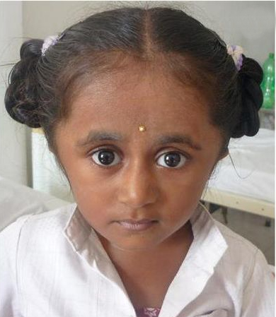
Musculoskeletal system
Assess for bone deformities, spinal anomalies, and joint hypermobility, with a focus on functional limitations caused by musculoskeletal abnormalities.
Cardiovascular system
Assess the heart rhythm and rate, and for murmurs. Assess for signs of arrhythmia, such as atrial fibrillation, and for signs of heart failure, as indicated by symptoms.
Cardiac valvular insufficiency, especially involving mitral and aortic valves, are well recognised features.[50]Folkestad L, Hald JD, Gram J, et al. Cardiovascular disease in patients with osteogenesis imperfecta - a nationwide, register-based cohort study. Int J Cardiol. 2016 Dec 15;225:250-7.
https://www.internationaljournalofcardiology.com/article/S0167-5273(16)32618-3/fulltext
http://www.ncbi.nlm.nih.gov/pubmed/27741483?tool=bestpractice.com
[56]Najib MQ, Schaff HV, Ganji J, et al. Valvular heart disease in patients with osteogenesis imperfecta. J Card Surg. 2013 Mar;28(2):139-43.
http://www.ncbi.nlm.nih.gov/pubmed/23347109?tool=bestpractice.com
One observational cohort study reported higher frequencies of heart failure, and atrial fibrillation or flutter in adults with OI compared with the general population.[50]Folkestad L, Hald JD, Gram J, et al. Cardiovascular disease in patients with osteogenesis imperfecta - a nationwide, register-based cohort study. Int J Cardiol. 2016 Dec 15;225:250-7.
https://www.internationaljournalofcardiology.com/article/S0167-5273(16)32618-3/fulltext
http://www.ncbi.nlm.nih.gov/pubmed/27741483?tool=bestpractice.com
Respiratory system
Assess for intrinsic and extrinsic causes of reduced pulmonary function, including assessment of:
Breath sounds
Chest wall appearance and expansion (e.g., assess for pectus excavatum and pectus carinatum)
Chest wall tenderness (may be indicative of rib fractures)
Spine curvature (scoliosis)
General mobility/immobility.
Skin
Assess for signs of skin fragility and easy bruising, which can commonly occur in OI.
In one study of 959 adults with OI, 42% self-reported a history of bruising.[28]Tosi LL, Oetgen ME, Floor MK, et al. Initial report of the osteogenesis imperfecta adult natural history initiative. Orphanet J Rare Dis. 2015 Nov 14;10:146.
https://ojrd.biomedcentral.com/articles/10.1186/s13023-015-0362-2
http://www.ncbi.nlm.nih.gov/pubmed/26578084?tool=bestpractice.com
Central nervous system
Assess for signs suggestive of basilar impression (sensory and motor deficits that suggest a cervical cord or cervical spinal root involvement).
Assess thoracic, lumbar, and sacral nerve root functions.
Initial investigations
A skeletal survey is recommended in most patients when establishing care and to assess the extent of fractures and bone deformities. A skeletal survey includes radiographs of skull; bilateral upper limbs including wrists and hands; bilateral lower limbs including pelvis and feet; anterior-posterior and lateral views of chest; and anterior-posterior and lateral views of cervical, thoracis, and lumbar spine.
Radiographic features will vary according to the type and severity of OI:[57]Weaver JS, Revels JW, Elifritz JM, et al. Clinical manifestations and medical imaging of osteogenesis imperfecta: fetal through adulthood. Acta Med Acad. 2021 Aug;50(2):277-91.
https://www.doi.org/10.5644/ama2006-124.343
http://www.ncbi.nlm.nih.gov/pubmed/34847680?tool=bestpractice.com
[58]Shapiro JR, Lietman C, Grover M, et al. Phenotypic variability of osteogenesis imperfecta type V caused by an IFITM5 mutation. J Bone Miner Res. 2013 Jul;28(7):1523-30.
https://academic.oup.com/jbmr/article/28/7/1523/7598577
http://www.ncbi.nlm.nih.gov/pubmed/23408678?tool=bestpractice.com
[59]Semler O, Cheung MS, Glorieux FH, et al. Wormian bones in osteogenesis imperfecta: correlation to clinical findings and genotype. Am J Med Genet A. 2010 Jul;152A(7):1681-7.
http://www.ncbi.nlm.nih.gov/pubmed/20583157?tool=bestpractice.com
People with OI type I and OI type IV may have wormian bones (small, irregular bones/ossicles that develop from extra ossification centres within the cranium) on the skull, vertebral wedging, and thin cortices of long bones
Children with OI type III may have flared metaphyses with popcorn calcification (radiologic appearance of calcification with irregular rings and arc, resembling popcorn)
People with OI type V may have hyperplastic callus and calcification of interosseous membranes
Most people with severe forms of OI have evidence of multiple bone deformities and kyphoscoliosis.
Consider referring all patients in whom diagnosis is to be established for genetic testing. Pretest counselling and interpretation of genetic test results should be done by medical geneticists and/or genetic counsellors. Genetic tests available for testing include:
Massively parallel (also known as next generation) sequencing-based gene panel testing for OI or skeletal dysplasia
Sanger sequencing for COL1A1 and COL1A2 (limited availability as this test has been largely superseded by massively parallel sequencing-based gene panel testing)
Exome sequencing
Whole genome sequencing.
Genetic testing can detect variants that are categorised by diagnostic laboratories into the following categories:
Note that a negative test may not always exclude OI; seek specialist input if there is diagnostic doubt.
Request dual-energy x-ray absorptiometry (DXA) in all (age-appropriate) patients when establishing care to help quantify changes in bone mineral density over time as well as to assess response to treatment.[60]Bianchi ML, Leonard MB, Bechtold S, et al. Bone health in children and adolescents with chronic diseases that may affect the skeleton: the 2013 ISCD Pediatric Official Positions. J Clin Densitom. 2014 Apr-Jun;17(2):281-94.
http://www.ncbi.nlm.nih.gov/pubmed/24656723?tool=bestpractice.com
[61]Liu W, Lee B, Nagamani SCS, et al. Approach to the patient: pharmacological therapies for fracture risk reduction in adults with osteogenesis imperfecta. J Clin Endocrinol Metab. 2023 Jun 16;108(7):1787-96.
https://www.ncbi.nlm.nih.gov/pmc/articles/PMC10271227
http://www.ncbi.nlm.nih.gov/pubmed/36658750?tool=bestpractice.com
This imaging modality is used to measure a 2-dimensional (areal) bone mineral content and bone mineral density.
People with OI typically have very low areal bone mineral density compared with age-matched peers in children and young adults and compared with peak bone mass in older adults.[8]Patel RM, Nagamani SC, Cuthbertson D, et al. A cross-sectional multicenter study of osteogenesis imperfecta in North America - results from the linked clinical research centers. Clin Genet. 2015 Feb;87(2):133-40.
https://www.ncbi.nlm.nih.gov/pmc/articles/PMC5529599
http://www.ncbi.nlm.nih.gov/pubmed/24754836?tool=bestpractice.com
[62]Bains JS, Carter EM, Citron KP, et al. A multicenter observational cohort study to evaluate the effects of bisphosphonate exposure on bone mineral density and other health outcomes in osteogenesis imperfecta. JBMR Plus. 2019 May;3(5):e10118.
https://www.ncbi.nlm.nih.gov/pmc/articles/PMC6524673
http://www.ncbi.nlm.nih.gov/pubmed/31131341?tool=bestpractice.com
DXA should not be performed in a child if safe and appropriate positioning cannot be assured.[60]Bianchi ML, Leonard MB, Bechtold S, et al. Bone health in children and adolescents with chronic diseases that may affect the skeleton: the 2013 ISCD Pediatric Official Positions. J Clin Densitom. 2014 Apr-Jun;17(2):281-94.
http://www.ncbi.nlm.nih.gov/pubmed/24656723?tool=bestpractice.com
Other investigations
It is recommended, based on experience in practice, that:
Baseline audiology evaluation is performed in all patients at diagnosis; the frequency of further audiological evaluations should be based on the severity of hearing loss, with particular consideration given to the impact hearing loss on quality of life for the patient.
Hearing evaluation is performed in children with OI before they start school and repeated every 3 years; annual evaluation should be carried out if abnormalities in hearing are detected.
Adults with reported hearing loss should have annual testing and follow-up appointments similar to the schedule for children.
If hearing loss is identified, regular ENT/audiology follow-up is recommended, with further review if hearing changes.
Adults experiencing tinnitus or symptoms of hearing loss should also have an audiological assessment to determine if hearing loss is conductive or sensorineural.
One study carried out in 133 patients with OI found that 57.9% had hearing loss on audiometry; hearing loss was progressive, often of mixed type, mostly bilateral, and began in the second to fourth decades of life.[40]Kuurila K, Kaitila I, Johansson R, et al. Hearing loss in Finnish adults with osteogenesis imperfecta: a nationwide survey. Ann Otol Rhinol Laryngol. 2002 Oct;111(10):939-46.
http://www.ncbi.nlm.nih.gov/pubmed/12389865?tool=bestpractice.com
Consider spirometry examination in patients when establishing care, to evaluate lung function in OI, and especially in patients with severe OI and those with scoliosis and chest deformities.[45]Chaney H, Mekking D, De Bakker D, et al. Key4OI recommendations for lung function guidance in osteogenesis imperfecta: based on an internationally performed comprehensive international consortium for health outcomes measurement procedure. Chest. 2023 May;163(5):1201-13.
https://journal.chestnet.org/article/S0012-3692(23)00039-9/fulltext
http://www.ncbi.nlm.nih.gov/pubmed/36640996?tool=bestpractice.com
On spirometry, forced vital capacity and FEV₁ are reduced in people with OI, with significantly reduced lung volumes in those with OI type III.[63]Tam A, Chen S, Schauer E, et al. A multicenter study to evaluate pulmonary function in osteogenesis imperfecta. Clin Genet. 2018 Dec;94(6):502-11.
https://www.ncbi.nlm.nih.gov/pmc/articles/PMC6235719
http://www.ncbi.nlm.nih.gov/pubmed/30152014?tool=bestpractice.com
Standing height is commonly used to estimate predictive values for many indices used in evaluating pulmonary function; be aware, however, that people with OI typically have a lower than average standing height, which may result in overestimation of pulmonary function status.[64]Sylvester KP, Clayton N, Cliff I, et al. ARTP statement on pulmonary function testing 2020. BMJ Open Respir Res. 2020 Jul;7(1):e000575.
https://bmjopenrespres.bmj.com/content/7/1/e000575.long
http://www.ncbi.nlm.nih.gov/pubmed/32631927?tool=bestpractice.com
Periodic evaluations are recommended in individuals with compromised lung functions.[63]Tam A, Chen S, Schauer E, et al. A multicenter study to evaluate pulmonary function in osteogenesis imperfecta. Clin Genet. 2018 Dec;94(6):502-11.
https://www.ncbi.nlm.nih.gov/pmc/articles/PMC6235719
http://www.ncbi.nlm.nih.gov/pubmed/30152014?tool=bestpractice.com
Request an echocardiogram in people with symptoms suggestive of cardiovascular disease and in those with physical findings suggestive of valvular disease.