Aetiology
Popliteal cyst formation has been attributed to underlying knee pathology, including trauma, arthritis, and infection (septic arthritis). Such aetiologies result in the development of knee joint effusion, which leads to popliteal cyst formation.
Injury of the medial meniscus, chondral lesions, and tears of the anterior cruciate ligament are the most commonly reported traumatic causes.[5][10] Ninety-four percent of popliteal cysts studied have been associated with intra-articular lesions.[5] Meniscal tear may be the most common intra-articular lesion: in one study, they were identified in 170 of 238 knees (71%) with a popliteal cyst.[10] A subsequent report noted a stronger correlation with chondral lesions.[11]
Arthritic causes associated with popliteal cysts include rheumatoid arthritis and osteoarthritis.[6][7] In 99 unselected consecutive patients with rheumatoid arthritis who underwent clinical and laboratory evaluation with knee radiographs and ultrasound of both knees, a popliteal cyst was detected in 47 patients (47.5%).[6] A statistically significant correlation was found between the presence of a popliteal cyst and clinical and radiological involvement of the knee by rheumatoid arthritis.
Joint infection with tuberculosis, coccidioidomycosis, candidiasis, or brucellosis may present with a popliteal cyst.[12][13][14][15] These joint infections result in synovitis and increased intra-articular pressures.
Another cause of popliteal cyst is pigmented villonodular synovitis.[16]
Pathophysiology
The synovium lines intra-articular joints. It is made up of a fibrillar interstitial matrix containing synovial cells. A rich meshwork of fenestrated microvessels in the synovial capsule produces the synovial fluid. Synovial fluid is formed continuously from a physiological osmotic gradient between the synovial microvasculature and the intra-articular space.[17]
Knee pathologies such as trauma, arthritis, or infection may result in increased synovial fluid volume and pressure.[17][18] Effusions develop when the absorption of synovial fluid lags behind microvascular filtration. Intra-articular pressure increases significantly during knee extension, and this may explain why pain occurs most often in this position. Although many reports have suggested an inherent weakness in the posterior capsule, no data support this.[Figure caption and citation for the preceding image starts]: Synovitis within cystFrom the collection of Dr John D. Kelly IV; used with permission [Citation ends].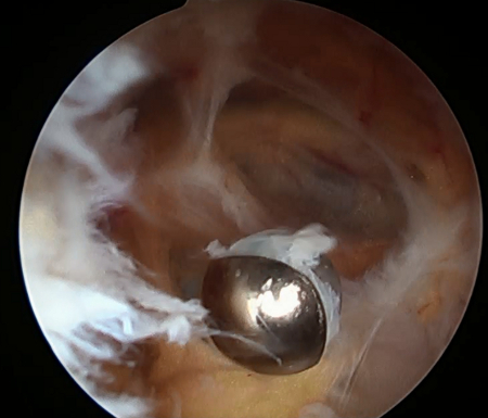
One study demonstrated that popliteal cysts develop from lack of external anatomical support.[1] This area is found between the two expansions of the semimembranosus muscle and the medial gastrocnemius.[Figure caption and citation for the preceding image starts]: MRI depicting cyst in interval of medial gastrocnemius and semimembranosusFrom the collection of Dr John D. Kelly IV; used with permission [Citation ends].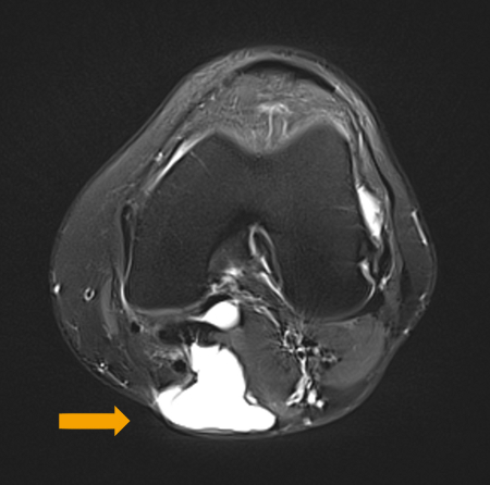 [Figure caption and citation for the preceding image starts]: Interval of medial gastrocnemius and semimembranosus viewed with arthroscopeFrom the collection of Dr John D. Kelly IV; used with permission [Citation ends].
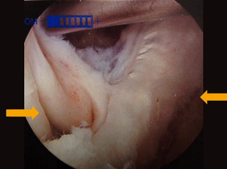
The posterior horn of the medial meniscus is the only reinforcing structure at this location. Degeneration of the medial meniscus, together with conditions where intra-articular pressure increases, makes the weak capsule protrude, giving rise to a popliteal cyst in the posteromedial popliteal fossa.[1][Figure caption and citation for the preceding image starts]: Preoperative T2-weighted MRI showing posterior fluid collection communicating with posterior medial joint in a patient with a popliteal cyst secondary to pigmented villonodular synovitisAdapted from Tosti R, Kelly JD 4th. Pigmented villonodular synovitis presenting as a Baker cyst. Am J Orthop (Belle Mead NJ). 2011;40:528-531; used with permission [Citation ends]. [Figure caption and citation for the preceding image starts]: Muscles surrounding the popliteal fossa (view of posterior aspect of right knee)Adapted by BMJ Evidence Centre from Gray's Anatomy [Citation ends].
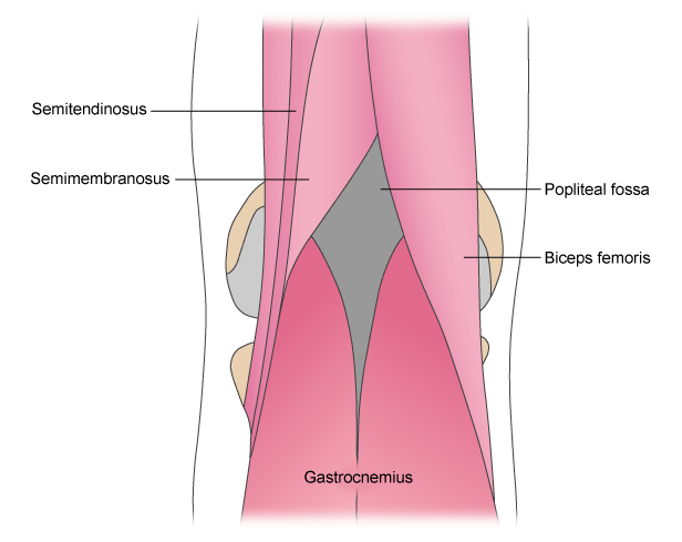
Use of this content is subject to our disclaimer