While technetium-99m bone scans can help to predict outcome, it usually takes 1 to 3 months to determine the prognosis.[4]McIntosh SE, Freer L, Grissom CK, et al. Wilderness Medical Society clinical practice guidelines for the prevention and treatment of frostbite: 2024 update. Wilderness Environ Med. 2024 Jun;35(2):183-97.
https://journals.sagepub.com/doi/10.1177/10806032231222359
http://www.ncbi.nlm.nih.gov/pubmed/38577729?tool=bestpractice.com
To determine prognosis, it is useful to classify injured tissue based on the topography of the lesion and early bone scan results, beginning at day 0 (just after rewarming).[1]Freer L, Handford C, Imray C. Frostbite. In: Auerbach PS. Wilderness medicine. 7th ed. Philadelphia, PA: Mosby Elsevier; 2017:197.
First- and second-degree injuries usually heal well and have a good prognosis. They do not typically require hospitalisation.
Third- and fourth-degree injuries may require amputation months later, after the viability of the tissue is determined. These patients often require hospitalisation and a full work-up because they are at increased risk of amputation and long-term sequelae, including decreased functionality of their limbs.[1]Freer L, Handford C, Imray C. Frostbite. In: Auerbach PS. Wilderness medicine. 7th ed. Philadelphia, PA: Mosby Elsevier; 2017:197.[3]Bhatnagar A, Sarker BB, Sawroop K, et al. Diagnosis, characterization and evaluation of treatment response of frostbite using pertechnetate scintigraphy: a prospective study. Eur J Nucl Med Mol Imaging. 2002 Feb;29(2):170-5.
http://www.ncbi.nlm.nih.gov/pubmed/11926378?tool=bestpractice.com
[9]Rabold MB. Frostbite and other localized cold-related injuries. In: Tintinalli JE, Kelen GD, Stapczynski JS, et al. Tintinalli's emergency medicine: a comprehensive study guide. 6th ed. New York, NY: McGraw-Hill; 2004.[14]Handford C, Buxton P, Russell K, et al. Frostbite: a practical approach to hospital management. Extrem Physiol Med. 2014 Apr 22;3:7.
https://www.ncbi.nlm.nih.gov/pmc/articles/PMC3994495
http://www.ncbi.nlm.nih.gov/pubmed/24764516?tool=bestpractice.com
Good clinical prognostic signs during the healing process are: early recovery of pinprick sensation; healthy-appearing skin; and the presence of clear, rather than haemorrhagic, blebs.[41]Zaramo TZ, Green JK, Janis JE. Practical review of the current management of frostbite injuries. Plast Reconstr Surg Glob Open. 2022 Oct 24;10(10):e4618.
https://www.ncbi.nlm.nih.gov/pmc/articles/PMC9592504
http://www.ncbi.nlm.nih.gov/pubmed/36299821?tool=bestpractice.com
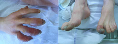
Poor clinical prognostic signs include: haemorrhagic blebs that do not extend distally, cyanosis, and tissue that appears frozen.[Figure caption and citation for the preceding image starts]: Typical frostbite injuries in the hands and feet of a climber with mildly haemorrhagic bullae presenting 3 days after exposure. The bullae were aseptically aspirated and a 5-day iloporost infusion resulted in a complete recoveryHallam M-J, BMJ 2010;341:c5864 [Citation ends].