Investigations
1st investigations to order
fluorescein angiogram for confirmation of diagnosis
Test
Occurs in affected region of retina: all major veins in central retinal vein occlusion (CRVO), 1 major or secondary vein in branch retinal vein occlusion (BRVO), superonasal and superotemporal branches or inferonasal and inferotemporal branches in hemiretinal vein occlusion (HRVO).
Areas of blocked fluorescence occur in regions of intra-retinal haemorrhage, which are present only in area affected by RVO.[Figure caption and citation for the preceding image starts]: Colour photograph, left eye; branch retinal vein occlusion; multiple intra-retinal images in quadrant of blocked veinFrom the personal library of Dr Aziz Khanifar [Citation ends].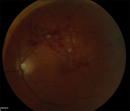 [Figure caption and citation for the preceding image starts]: Fluorescein angiogram, right eye; central retinal vein occlusion; delayed drainage of veins in each quadrantFrom the personal library of Dr Aziz Khanifar [Citation ends].
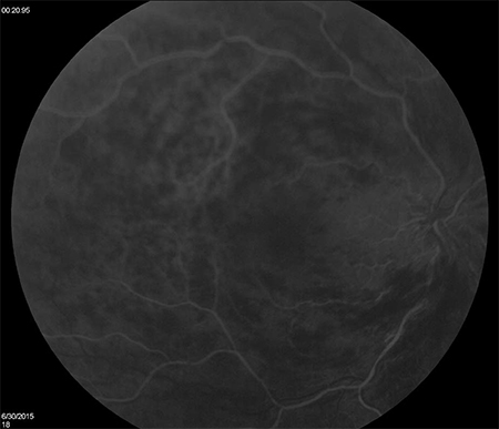
Result
delayed venous filling during transit phase; areas of blocked fluorescence
Investigations to consider
fluorescein angiogram for assessment of complications
Test
Areas of non-perfusion suggest significant ischaemia. In CRVO, the presence of 10 disc areas of non-perfusion suggests that the RVO is 'ischaemic'. In BRVO, the presence of 5 disc areas of non-perfusion suggests that the RVO is 'non-perfused'.
Petalloid parafoveal leakage suggests cystoid macular oedema. Can also emanate from collateral vessels in BRVO.
Areas of neovascularisation have significant leakage.
Result
areas of non-perfusion; petalloid parafoveal leakage; leakage from retinal neovascular complex
optical coherence tomography
Test
Confirms presence of macular oedema.
May reveal intraretinal hyporeflectivity and thickening of the neurosensory retina (evidence of cystoid macular oedema).[26][27] Sub-retinal hyporeflectivity (evidence of sub-retinal fluid) can be found in eyes with large amounts of macular oedema. Can be quantified in microns (retinal thickness) or as cubic millimetres (total macular volume). Can be used to evaluate response to therapy for macular oedema. Macular oedema will be present asymmetrically in a BRVO. For example, for a superior BRVO, only the superior half of the macula will be oedematous.[Figure caption and citation for the preceding image starts]: Fluorescein angiogram, left eye; branch retinal vein occlusion; delayed drainage of blocked vein superotemporallyFrom the personal library of Dr Aziz Khanifar [Citation ends].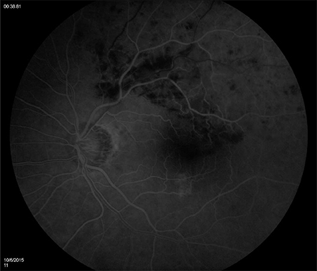
Result
cystoid retinal thickening in macular oedema
electroretinography
Test
Can confirm perfusion status of CRVO (especially in setting of vitreous haemorrhage or significant intra-retinal haemorrhage that prohibits utility of fluorescein angiogram).[25]
Very infrequently used in routine clinical practice.
Result
mean b-wave amplitude reduced by ≥40%
Use of this content is subject to our disclaimer