Approach
The only true treatment of melanocytic nevi is complete (as opposed to incomplete) removal of the lesion. This is recommended for accurate histopathological diagnosis and to reduce the risk of recurrence or persistence. Incompletely excised melanocytic nevi confer an increased risk of sampling error, and recurrent or persistent melanocytes may result in a pseudomelanoma pattern clinically and histologically, further confusing the diagnosis.[47][48][50][51] The incidence of recurrence is higher with shave excision, as the nevus is less likely to be completely excised, especially at depth.[32] Therefore, shave or saucerisation techniques are not recommended for the complete removal of nevi.
Skin procedures are generally well tolerated, with a small risk of bleeding, infection (usually <1%), haematoma, and nerve damage (usually sensory; rarely motor).[5] Allergy to local anaesthetic is rare, especially with the amide local anaesthetics such as lidocaine (vs. ester local anaesthetics). The most commonly used local anaesthetic is 1% lidocaine with a 1:100,000 dilution of adrenaline (epinephrine), which is added for its vasoconstrictive properties, helping to decrease bleeding and prolong the duration of anaesthesia. Any pain is usually minimal and easily managed with paracetamol. Randomised controlled trials have found no increase in infection rates or wound healing when comparing topical antibiotics and white soft paraffin (petroleum jelly) in clean dermatological wounds.[75][76] Topical antibiotics, neomycin and bacitracin being among the most frequent, are common sensitising agents in the development of allergic contact dermatitis, and may rarely cause anaphylaxis.[75][77] For these reasons, and for the additional benefit of cost-saving, white soft paraffin (petroleum jelly) or an emollient is generally recommended for wound care post-procedure. Hypoallergenic adhesive tape may also be substituted for sensitive skin if adhesives in adhesive bandages are irritating. The scar formed by the procedure may hypertrophy or become a keloid, and depending on location may be cosmetically or functionally undesirable.[Figure caption and citation for the preceding image starts]: A persistent or recurrent nevusFrom the collection of Jason Lee, Thomas Jefferson University [Citation ends].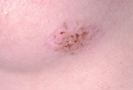
Clinically benign
If the physician is confident from history and physical examination that the melanocytic nevus is benign, then treatment (i.e., removal) is not usually indicated. The patient may require only reassurance of the benign nature of the nevus. Some patients, despite being reassured, may still want the nevus to be removed. This may be due to:
Repetitive trauma to the lesion
Symptoms such as itching, bleeding, or irritation
Cosmetic reasons
Excessive anxiety that the lesion is malignant despite reassurance.
Punch excision biopsy
If the lesion is small enough, it may fit within the bore of the trephine, allowing complete removal of the nevus with relatively small margins of safety. The advantage of this technique is a relatively small scar without having to remove redundant standing cones of tissue or use deep sutures. This technique would not be used if there were concern for the potential for malignancy.
Excision biopsy
Excision of the lesion is the definitive method for removal of melanocytic lesions.[5][46] A small margin of tissue (2 mm) is provided to ensure free margins, and the depth is removed down to the level of the fat, ensuring complete removal. Disadvantages of this method are that it is a more invasive procedure with a potentially increased risk of infection and surgical complications (such as bleeding and haematoma), and sutures are required, necessitating the patient's return within 1 to 2 weeks for removal.
Laser
A variety of laser devices are used for the treatment of various pigmented lesions, including some types of melanocytic nevi.[5][32][78][79] Although utilised by some, the practice is controversial, especially because of the lack of histopathological diagnosis and the potential for misdiagnosis of a melanoma as a benign nevus or lentigo.[48][80] The potential for malignant change of melanocytes treated by laser is unknown, and there have been case reports of melanomas arising in sites treated by laser.[81][82] Additionally, incomplete removal of the nevus may result in a pseudomelanoma pattern.[83] For these reasons, chemical peeling and other non-excisional methods (utilised mainly for giant congenital nevi) are not recommended.[48][49]
Clinically suspicious for melanoma
The most important reasons for removal of a melanocytic nevus are if there is high clinical suspicion of melanoma; if there is a history of change in the lesion, supported by physical examination; and/or if there is a high suspicion of atypical features suggestive of melanoma.[46] Dysplastic nevi are one kind of benign nevus that can be clinically difficult to distinguish from melanoma, but it is important to understand that they are a type of benign nevus and should not be indiscriminately biopsied.[17][32][47][48] Most melanomas arise de novo rather than in association with a nevus, so prophylactic removal of nevi is not warranted.[32] Biopsy should be reserved for lesions suggestive of malignant changes; removal of all dysplastic nevi is not recommended.[1][4][5][30][31][32][47][48] If the clinician is uncertain about the distinction between melanocytic nevus and melanoma, a dermatology referral should be considered.
Excisional biopsy is the preferred method for removing a melanocytic lesion that is clinically suspicious for melanoma.
Use of this content is subject to our disclaimer