Images and videos
Images


Superior vena cava syndrome
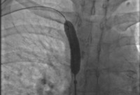
Stent deployment in the superior vena cava
Image obtained from cardiac catheterisation laboratory at University of Missouri, Columbia; used with permission
See this image in context in the following section/s:
Superior vena cava syndrome
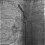
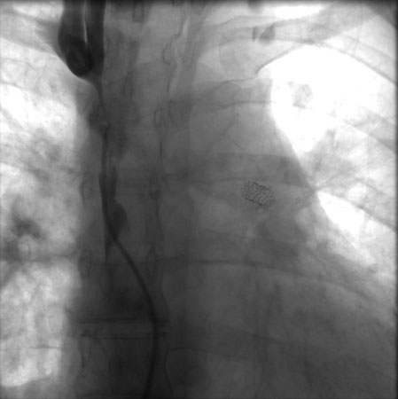
Venography showing superior vena cava stenosis. Stent placement in the left pulmonary artery is seen
Image obtained from cardiac catheterisation laboratory at University of Missouri, Columbia; used with permission
See this image in context in the following section/s:

Superior vena cava syndrome
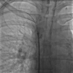
Post-dilatation of the superior vena cava stent
Image obtained from cardiac catheterisation laboratory at University of Missouri, Columbia; used with permission
See this image in context in the following section/s:

Superior vena cava syndrome
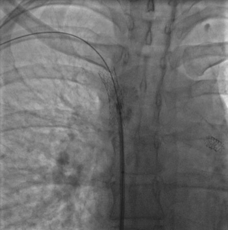
Percutaneous balloon angioplasty of the stenotic lesion in superior vena cava
Image obtained from cardiac catheterisation laboratory at University of Missouri, Columbia; used with permission
See this image in context in the following section/s:
Superior vena cava syndrome
Supra- and infra-azygos obstruction leading to superior vena cava (SVC) syndrome. IVC: inferior vena cava
Reproduced with permission from Braunwald's Heart Disease, 8th ed (2008)
See this image in context in the following section/s:
Use of this content is subject to our disclaimer