Aetiology
A total of 65% to 70% of cases are due to malignancy.[1][2] Lung cancer is the most common aetiology, with non-small cell lung cancer accounting for 50% of cases of malignant superior vena cava (SVC) syndrome and small cell lung cancer for 25% cases.[9][10][11] Overall, 2% to 5% of all patients with lung cancer go on to develop SVC syndrome, while 10% to 20% of patients with small cell lung cancer develop the condition. This is most probably related to the central growth of these tumours.[12] Most patients with lung cancer-associated SVC syndrome have right-sided lesions (80%). Lymphoma is the second most common cause, accounting for 12% of cases of malignant SVC syndrome; diffuse large cell lymphoma is the most common type (two-thirds), followed by lymphoblastic lymphoma (one third). Of patients with primary mediastinal B-cell lymphoma with sclerosis (a rare subtype of non-Hodgkin's lymphoma), the prevalence of SVC syndrome is high, with one study reporting 57%.[13] Although Hodgkin's lymphoma often involves the mediastinum, SVC syndrome is rare.[14] Thymoma (2%) and germ cell tumours (3%) are other primary mediastinal malignancies that occasionally cause SVC syndrome.[15]
The most common metastatic disease that causes SVC syndrome is breast cancer, accounting for 11% of cases.[6] Other metastatic tumours that cause SVC syndrome include colon cancer, oesophageal cancer, Kaposi's sarcoma, and fibrous mesothelioma.
Benign causes of SVC syndrome are less frequent (30%-35%).[1][2] They include iatrogenic causes associated with SVC thrombosis (e.g., central venous catheters, pacemaker and implantable cardioverter-defibrillator leads), mediastinal fibrosis caused by radiotherapy or infections (e.g., histoplasmosis, tuberculosis, aspergillosis, blastomycosis, or nocardiosis), collagen-vascular diseases like sarcoidosis or Behcet's syndrome, and, rarely, aortic arch aneurysm, large substernal goitre, mediastinal haematoma as a result of trauma, and bicaval anastomotic stenosis following cardiac transplantation.[1][11][16] Complications of pacemaker lead placement, such as venous thrombosis and stenosis, occur in up to 30% of patients, but only a few patients become symptomatic; however, the presence of multiple leads, retention of severed lead(s), and previous lead infection may increase risk of SVC syndrome.
In children, SVC syndrome is most often caused by non-Hodgkin's lymphoma. The compression of the SVC may be associated with compression of the trachea, which is narrow, flexible, and soft relative to that of an adult. This may result in airway obstruction in children.
Pathophysiology
The superior vena cava (SVC) extends from the junction of the right and left brachiocephalic veins to the right atrium. It drains venous blood from the head, neck, upper extremities, and upper thorax to the right atrium. It is located in the middle mediastinum and is surrounded by structures including the trachea, right bronchus, aorta, pulmonary artery, and the perihilar and paratracheal lymph nodes.
The thin-walled SVC can be obstructed by intraluminal, mural, or extrinsic factors. The extent and rapidity of obstruction correlates with elevation of venous pressure and symptomatic presentation. Slowly progressive obstruction leads to recruitment of collateral circulation via the azygos and internal mammary venous system (which takes several weeks).[17] With obstruction of the SVC, the cervical venous pressure is usually increased to 20 to 40 mmHg (normal range 2-8 mmHg).[18] When obstruction occurs abruptly, SVC syndrome can constitute a medical emergency.
An obstructed SVC initiates collateral venous return to the heart from the upper half of the body through four principal pathways. The most important pathway is the azygos venous system, which includes the azygos vein, the hemiazygos vein, and the connecting intercostal veins. The second pathway is the internal mammary venous system plus tributaries and secondary communications to the superior and inferior epigastric veins. The long thoracic venous system, with its connections to the femoral veins and vertebral veins, provides the third and fourth collateral routes, respectively.[1]
Classification
Classification based on location of superior vena cava (SVC) obstruction
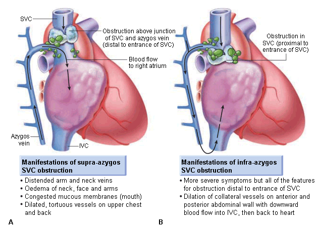
Pre-azygos or supra-azygos
Obstruction of blood return above the entrance of azygos vein into the SVC, resulting in venous distension and oedema of the face, neck, and upper extremities.[3][4]
Post-azygos or infra-azygos
Obstruction below the entrance of azygos vein into the SVC results in retrograde flow through the azygos via collaterals to the inferior vena cava, resulting in not only the symptoms and signs of pre-azygos disease, but also dilation of the veins over the abdomen.
This is usually more severe and poorly tolerated than pre-azygos obstruction. [Figure caption and citation for the preceding image starts]: Supra- and infra-azygos obstruction leading to superior vena cava (SVC) syndrome. IVC: inferior vena cavaReproduced with permission from Braunwald's Heart Disease, 8th ed (2008) [Citation ends].

Classification based on aetiology of obstruction
Luminal obstruction (e.g., pacemaker leads or catheter-related thrombosis).
Extrinsic compression (e.g., malignancy, fibrosing mediastinitis due to infection/radiation, aortic arch aneurysm, haematoma, or goitre).
Use of this content is subject to our disclaimer