The initial assessment of suspected asthma focuses on the presence of key features in the history, clinical examination, and medical records, together with careful consideration of risk factors and alternative diagnoses.[1]Global Initiative for Asthma. 2024 Global strategy for asthma management and prevention. May 2024 [internet publication].
https://ginasthma.org/wp-content/uploads/2024/05/GINA-2024-Strategy-Report-24_05_22_WMS.pdf
[8]National Institute for Health and Care Excellence. Asthma: diagnosis, monitoring and chronic asthma management (BTS, NICE, SIGN). Nov 2024 [internet publication].
https://www.nice.org.uk/guidance/ng245
[97]Moral L, Vizmanos G, Torres-Borrego J, et al. Asthma diagnosis in infants and preschool children: a systematic review of clinical guidelines. Allergol Immunopathol (Madr). 2019 Mar-Apr;47(2):107-21.
http://www.ncbi.nlm.nih.gov/pubmed/30193886?tool=bestpractice.com
[98]National Institute for Health and Care Excellence. Asthma pathway (BTS, NICE, SIGN). Nov 2024 [internet publication].
https://www.nice.org.uk/guidance/ng244
Diagnosis is age-dependent and requires a history of variable expiratory airflow limitation, in addition to symptom improvement with a clinical trial of inhaled bronchodilators and corticosteroids.[1]Global Initiative for Asthma. 2024 Global strategy for asthma management and prevention. May 2024 [internet publication].
https://ginasthma.org/wp-content/uploads/2024/05/GINA-2024-Strategy-Report-24_05_22_WMS.pdf
[8]National Institute for Health and Care Excellence. Asthma: diagnosis, monitoring and chronic asthma management (BTS, NICE, SIGN). Nov 2024 [internet publication].
https://www.nice.org.uk/guidance/ng245
The basis of an asthma diagnosis or treatment decision should be clearly documented.[1]Global Initiative for Asthma. 2024 Global strategy for asthma management and prevention. May 2024 [internet publication].
https://ginasthma.org/wp-content/uploads/2024/05/GINA-2024-Strategy-Report-24_05_22_WMS.pdf
[8]National Institute for Health and Care Excellence. Asthma: diagnosis, monitoring and chronic asthma management (BTS, NICE, SIGN). Nov 2024 [internet publication].
https://www.nice.org.uk/guidance/ng245
See Criteria.
Treat children immediately if they are acutely unwell or highly symptomatic at presentation.[1]Global Initiative for Asthma. 2024 Global strategy for asthma management and prevention. May 2024 [internet publication].
https://ginasthma.org/wp-content/uploads/2024/05/GINA-2024-Strategy-Report-24_05_22_WMS.pdf
[8]National Institute for Health and Care Excellence. Asthma: diagnosis, monitoring and chronic asthma management (BTS, NICE, SIGN). Nov 2024 [internet publication].
https://www.nice.org.uk/guidance/ng245
[98]National Institute for Health and Care Excellence. Asthma pathway (BTS, NICE, SIGN). Nov 2024 [internet publication].
https://www.nice.org.uk/guidance/ng244
Diagnostic testing can be attempted, but consider waiting until the child is more stable. See Acute asthma exacerbation in children for more information about the diagnosis of acute exacerbations.
Probability-based approach to diagnosis
A probability-based approach plus a therapeutic trial is recommended for children younger than 5 years because most will be unable to perform lung function tests reliably; from age 5 years, children should have their asthma diagnosis confirmed with a test of variable expiratory airflow limitation.[1]Global Initiative for Asthma. 2024 Global strategy for asthma management and prevention. May 2024 [internet publication].
https://ginasthma.org/wp-content/uploads/2024/05/GINA-2024-Strategy-Report-24_05_22_WMS.pdf
[8]National Institute for Health and Care Excellence. Asthma: diagnosis, monitoring and chronic asthma management (BTS, NICE, SIGN). Nov 2024 [internet publication].
https://www.nice.org.uk/guidance/ng245
[99]Gupta S, Thériault G. Do not diagnose or routinely treat asthma or chronic obstructive pulmonary disease without pulmonary function testing. BMJ. 2023 Mar 20;380:e072834.
http://www.ncbi.nlm.nih.gov/pubmed/36940980?tool=bestpractice.com
If they cannot perform objective tests on one occasion, retry every 6-12 months until reliable results are obtained.[8]National Institute for Health and Care Excellence. Asthma: diagnosis, monitoring and chronic asthma management (BTS, NICE, SIGN). Nov 2024 [internet publication].
https://www.nice.org.uk/guidance/ng245
Although the peak expiratory flow (PEF) is less reliable than spirometry, its use is recommended where diagnosis would otherwise rely on symptoms alone.[1]Global Initiative for Asthma. 2024 Global strategy for asthma management and prevention. May 2024 [internet publication].
https://ginasthma.org/wp-content/uploads/2024/05/GINA-2024-Strategy-Report-24_05_22_WMS.pdf
[8]National Institute for Health and Care Excellence. Asthma: diagnosis, monitoring and chronic asthma management (BTS, NICE, SIGN). Nov 2024 [internet publication].
https://www.nice.org.uk/guidance/ng245
The following probability-based approach to diagnosis is proposed.[100]National Heart, Lung, and Blood Institute; National Asthma Education and Prevention Program. Guidelines for the diagnosis and management of asthma. Aug 2007 [internet publication].
http://www.nhlbi.nih.gov/health-pro/guidelines/current/asthma-guidelines/full-report.htm
High probability of asthma: start with a therapeutic trial and reserve further testing for those with a poor response.
Intermediate probability of asthma (can perform spirometry and has evidence of airway obstruction): offer a reversibility test and/or time-limited therapeutic trial.
If there is reversibility, or if treatment is beneficial, treat as asthma.
If there is no significant reversibility, and/or a therapeutic trial is not beneficial, consider tests for alternative conditions.
Intermediate probability of asthma (can perform spirometry and with no evidence of airway obstruction): consider testing for atopic status, bronchodilator reversibility, and, if possible, bronchial hyper-responsiveness using methacholine or exercise.
Intermediate probability of asthma (unable to perform spirometry): consider testing for atopic status and offering a time-limited therapeutic trial.
If beneficial, treat as asthma.
If not beneficial, stop treatment and consider an alternative diagnosis and/or consultant referral.
The greater the variations in expiratory lung function, and the more often excess variation is observed, the more confident a clinician can be with the diagnosis of childhood asthma.[1]Global Initiative for Asthma. 2024 Global strategy for asthma management and prevention. May 2024 [internet publication].
https://ginasthma.org/wp-content/uploads/2024/05/GINA-2024-Strategy-Report-24_05_22_WMS.pdf
However, it is important to ensure that apparent variability does not reflect variations in technique over time, because both spirometry and PEF measures are effort-dependent.
Do not confirm a diagnosis of asthma without a suggestive clinical history and a supporting objective test; instead, manage as suspected asthma until the diagnosis is confirmed. If objective tests cannot be done at presentation because the patient is very symptomatic, delay testing until the patient has stabilised on treatment. Once the diagnosis of asthma is confirmed, record the basis for diagnosis in the person's medical records.[1]Global Initiative for Asthma. 2024 Global strategy for asthma management and prevention. May 2024 [internet publication].
https://ginasthma.org/wp-content/uploads/2024/05/GINA-2024-Strategy-Report-24_05_22_WMS.pdf
[8]National Institute for Health and Care Excellence. Asthma: diagnosis, monitoring and chronic asthma management (BTS, NICE, SIGN). Nov 2024 [internet publication].
https://www.nice.org.uk/guidance/ng245
Using a structured questionnaire can help standardise this approach, but reliance on asthma prediction tools and screening aids is not recommended because they show wide variation in performance when assessing future risk.[101]Chang TS, Lemanske RF Jr, Guilbert TW, et al. Evaluation of the modified asthma predictive index in high-risk preschool children. J Allergy Clin Immunol Pract. 2013 Mar;1(2):152-6.
https://www.ncbi.nlm.nih.gov/pmc/articles/PMC3811153
http://www.ncbi.nlm.nih.gov/pubmed/24187656?tool=bestpractice.com
[102]Colicino S, Munblit D, Minelli C, et al. Validation of childhood asthma predictive tools: A systematic review. Clin Exp Allergy. 2019 Apr;49(4):410-8.
http://www.ncbi.nlm.nih.gov/pubmed/30657220?tool=bestpractice.com
[103]Kothalawala DM, Kadalayil L, Weiss VBN, et al. Prediction models for childhood asthma: a systematic review. Pediatr Allergy Immunol. 2020 Aug;31(6):616-27.
http://www.ncbi.nlm.nih.gov/pubmed/32181536?tool=bestpractice.com
[104]Daines L, McLean S, Buelo A, et al. Systematic review of clinical prediction models to support the diagnosis of asthma in primary care. NPJ Prim Care Respir Med. 2019 May 9;29(1):19.
http://www.ncbi.nlm.nih.gov/pubmed/31073125?tool=bestpractice.com
The Childhood Asthma Risk Tool (CHART) has been proposed; its performance in clinical care remains unclear.[105]Reyna ME, Dai R, Tran MM, et al. Development of a symptom-based tool for screening of children at high risk of preschool asthma. JAMA Netw Open. 2022 Oct 3;5(10):e2234714.
http://www.ncbi.nlm.nih.gov/pubmed/36201211?tool=bestpractice.com
Refer preschool children to a consultant respiratory paediatrician if they require hospital admission or ≥2 accident and emergency department admissions with wheeze in a 12-month period.[8]National Institute for Health and Care Excellence. Asthma: diagnosis, monitoring and chronic asthma management (BTS, NICE, SIGN). Nov 2024 [internet publication].
https://www.nice.org.uk/guidance/ng245
History
Recurrent symptoms of wheezing, cough (worse at night or early morning), and shortness of breath in response to recognised triggers (e.g., temperature change, viral infection, exercise, and emotion) are characteristic of asthma. Parental perception should be checked because various respiratory noises may be incorrectly labelled as wheezing (where possible, wheeze should be confirmed by a healthcare professional).[106]Cane RS, Ranganathan SC, McKenzie SA. What do parents of wheezy children understand by "wheeze"? Arch Dis Child. 2000 Apr;82(4):327-32.
https://adc.bmj.com/content/82/4/327.long
http://www.ncbi.nlm.nih.gov/pubmed/10735844?tool=bestpractice.com
The diagnosis is supported by features of atopic disease, such as eczema, atopic dermatitis, and allergic rhinitis in the child or first-degree family members.
Features associated with asthma include:
Episodic symptoms (wheeze, breathlessness, chest tightness, and cough occurring episodically with periods of no or minimal symptoms)
Wheeze confirmed by a healthcare professional
Diurnal variability (worse at night or early morning)
A history of atopy
Recurrent events over time
Cough is often misdiagnosed as asthma in children and requires a careful review of the history and the exclusion of alternative causes (see Differentials).[107]Lai K, Satia I, Song WJ, et al. Cough and cough hypersensitivity as treatable traits of asthma. Lancet Respir Med. 2023 Jul;11(7):650-62.
http://www.ncbi.nlm.nih.gov/pubmed/37336227?tool=bestpractice.com
Many children younger than 5 years present with recurrent wheezing due to frequent upper respiratory tract infections (URTIs).[1]Global Initiative for Asthma. 2024 Global strategy for asthma management and prevention. May 2024 [internet publication].
https://ginasthma.org/wp-content/uploads/2024/05/GINA-2024-Strategy-Report-24_05_22_WMS.pdf
Wheeze is a heterogeneous phenotype in young children, and many non-atopic children who experience recurrent episodes of viral-induced wheezing will not require a regular inhaled corticosteroids (ICS) or go on to develop chronic atopic asthma.
Physical examination
Most children have no signs when they do not have an exacerbation. Depending on the symptom pattern, examination may uncover widespread polyphonic wheeze audible on chest auscultation and respiratory distress (e.g., tachypnoea, recessions or retractions, and accessory muscle use). In poorly controlled persistent asthma, hyperinflation (reflecting gas trapping) and chest wall deformity (Harrison's sulci) may be present. Features of atopic disease may be evident on examination.
Initial tests
Chest x-ray and a full blood count (FBC) with differential are indicated to exclude other pathologies in patients presenting for the first time or with an acute exacerbation.
Chest imaging (x-ray or high-resolution computed tomography [CT]) may demonstrate hyperinflation in asthma, can diagnose bronchiectasis and situs inversus, and can distinguish cardiac from pulmonary diseases. Imaging is not recommended routinely to predict treatment outcomes or lung function or to assess treatment response.
FBC with differential may demonstrate eosinophilia or infection.
In the UK, joint British Thoracic Society, National Institute for Health and Care Excellence, and Scottish Intercollegiate Guidelines Network (BTS/NICE/SIGN) guidance recommends measuring the fractional exhaled nitric oxide (FeNO) level in children with a history suggestive of asthma and that asthma can be diagnosed if the FeNO level is ≥35 parts per billion (ppb).[8]National Institute for Health and Care Excellence. Asthma: diagnosis, monitoring and chronic asthma management (BTS, NICE, SIGN). Nov 2024 [internet publication].
https://www.nice.org.uk/guidance/ng245
However, this recommendation is specific to BTS/NICE/SIGN and is not currently recommended by other international guidelines.
See Acute asthma exacerbation in children.
Response to medication
Response to a therapeutic trial of inhaled beta-2 agonist or corticosteroid, given as either a short course of oral corticosteroid (e.g., 1-2 mg/kg/day for 3 days) or a longer trial of low-dose inhaled corticosteroid (e.g., for 4-6 weeks), is suggestive of asthma.[1]Global Initiative for Asthma. 2024 Global strategy for asthma management and prevention. May 2024 [internet publication].
https://ginasthma.org/wp-content/uploads/2024/05/GINA-2024-Strategy-Report-24_05_22_WMS.pdf
[8]National Institute for Health and Care Excellence. Asthma: diagnosis, monitoring and chronic asthma management (BTS, NICE, SIGN). Nov 2024 [internet publication].
https://www.nice.org.uk/guidance/ng245
Any therapeutic trial should be time-limited:
Consider a diagnosis of asthma if there is clinical improvement (based on symptom control and exacerbation rate) during treatment and deterioration when treatment is stopped.
Consider alternative diagnoses if there is a lack of response; cough and wheeze often have different causes in children than in adults, necessitating care to ensure proper investigation.[107]Lai K, Satia I, Song WJ, et al. Cough and cough hypersensitivity as treatable traits of asthma. Lancet Respir Med. 2023 Jul;11(7):650-62.
http://www.ncbi.nlm.nih.gov/pubmed/37336227?tool=bestpractice.com
This approach is also suitable for children with a non-specific cough and risk factors for asthma.[107]Lai K, Satia I, Song WJ, et al. Cough and cough hypersensitivity as treatable traits of asthma. Lancet Respir Med. 2023 Jul;11(7):650-62.
http://www.ncbi.nlm.nih.gov/pubmed/37336227?tool=bestpractice.com
[108]Morice AH, Millqvist E, Bieksiene K, et al. ERS guidelines on the diagnosis and treatment of chronic cough in adults and children. Eur Respir J. 2020 Jan;55(1):1901136.
http://www.ncbi.nlm.nih.gov/pubmed/31515408?tool=bestpractice.com
[109]Chang AB, Oppenheimer JJ, Irwin RS, et al. Managing chronic cough as a symptom in children and management algorithms: CHEST guideline and expert panel report. Chest. 2020 Jul;158(1):303-29.
http://www.ncbi.nlm.nih.gov/pubmed/32179109?tool=bestpractice.com
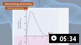
Spirometry
Spirometry is the preferred method for assessing variability in expiratory lung function. It is performed in children with suspected asthma who are able to give repeatable and reproducible results, but this is highly dependent on local service availability and the child's level of cooperation (typically from age 5 years).[1]Global Initiative for Asthma. 2024 Global strategy for asthma management and prevention. May 2024 [internet publication].
https://ginasthma.org/wp-content/uploads/2024/05/GINA-2024-Strategy-Report-24_05_22_WMS.pdf
[110]Gaillard EA, Kuehni CE, Turner S, et al. European Respiratory Society clinical practice guidelines for the diagnosis of asthma in children aged 5-16 years. Eur Respir J. 2021 Oct;58(5):2004173.
https://www.doi.org/10.1183/13993003.04173-2020
http://www.ncbi.nlm.nih.gov/pubmed/33863747?tool=bestpractice.com
The European Respiratory Society and American Thoracic Society (ERS/ATS) have jointly published standardised guidance for performing and interpreting spirometry.[111]Miller MR, Crapo R, Hankinson J, et al. General considerations for lung function testing. Eur Respir J. 2005 Jul;26(1):153-61.
http://www.ncbi.nlm.nih.gov/pubmed/15994402?tool=bestpractice.com
[112]Stanojevic S, Kaminsky DA, Miller MR, et al. ERS/ATS technical standard on interpretive strategies for routine lung function tests. Eur Respir J. 2022 Jul 13;60(1):2101499.
https://publications.ersnet.org/content/erj/60/1/2101499
http://www.ncbi.nlm.nih.gov/pubmed/34949706?tool=bestpractice.com
There are three core spirometry measurements:
Forced expiratory volume in 1 second (FEV₁): the total volume of air forcibly exhaled in the first second after one breath. Similar to the PEF.
Forced vital capacity (FVC): the total volume of air forcibly exhaled after one breath.
FEV₁/FVC: the ratio of FEV1 to FVC expressed as a percentage.
An obstructive pattern may be present, suggested by visual scalloping of the expiratory flow-volume loop. Decreases can be observed in the FEV₁/FVC ratio, FEV₁, or mid-flows (maximal expiratory flow at 25% of FVC [MEF25] or forced expiratory flow between 25% and 75% of FVC [FEF25-75]). The FEV₁/FVC ratio is normally >0.90 in children.[1]Global Initiative for Asthma. 2024 Global strategy for asthma management and prevention. May 2024 [internet publication].
https://ginasthma.org/wp-content/uploads/2024/05/GINA-2024-Strategy-Report-24_05_22_WMS.pdf
FEV₁ and FEV₁/FVC results below the lower limit of normal (LLN) or below 80% of the predicted value are generally considered suggestive of an asthma diagnosis.[1]Global Initiative for Asthma. 2024 Global strategy for asthma management and prevention. May 2024 [internet publication].
https://ginasthma.org/wp-content/uploads/2024/05/GINA-2024-Strategy-Report-24_05_22_WMS.pdf
[110]Gaillard EA, Kuehni CE, Turner S, et al. European Respiratory Society clinical practice guidelines for the diagnosis of asthma in children aged 5-16 years. Eur Respir J. 2021 Oct;58(5):2004173.
https://www.doi.org/10.1183/13993003.04173-2020
http://www.ncbi.nlm.nih.gov/pubmed/33863747?tool=bestpractice.com
The LLNs for spirometry values are age-, height-, and ethnicity-dependent.[110]Gaillard EA, Kuehni CE, Turner S, et al. European Respiratory Society clinical practice guidelines for the diagnosis of asthma in children aged 5-16 years. Eur Respir J. 2021 Oct;58(5):2004173.
https://www.doi.org/10.1183/13993003.04173-2020
http://www.ncbi.nlm.nih.gov/pubmed/33863747?tool=bestpractice.com
[111]Miller MR, Crapo R, Hankinson J, et al. General considerations for lung function testing. Eur Respir J. 2005 Jul;26(1):153-61.
http://www.ncbi.nlm.nih.gov/pubmed/15994402?tool=bestpractice.com
[112]Stanojevic S, Kaminsky DA, Miller MR, et al. ERS/ATS technical standard on interpretive strategies for routine lung function tests. Eur Respir J. 2022 Jul 13;60(1):2101499.
https://publications.ersnet.org/content/erj/60/1/2101499
http://www.ncbi.nlm.nih.gov/pubmed/34949706?tool=bestpractice.com
False normal FEV₁/FVC ratios are possible with an incorrect technique, irrespective of age, and normal spirometry results do not automatically exclude asthma.[1]Global Initiative for Asthma. 2024 Global strategy for asthma management and prevention. May 2024 [internet publication].
https://ginasthma.org/wp-content/uploads/2024/05/GINA-2024-Strategy-Report-24_05_22_WMS.pdf
[110]Gaillard EA, Kuehni CE, Turner S, et al. European Respiratory Society clinical practice guidelines for the diagnosis of asthma in children aged 5-16 years. Eur Respir J. 2021 Oct;58(5):2004173.
https://www.doi.org/10.1183/13993003.04173-2020
http://www.ncbi.nlm.nih.gov/pubmed/33863747?tool=bestpractice.com
Response to a beta-2 agonist bronchodilator (≥12% improvement from baseline FEV₁) is also suggestive of an asthma diagnosis.[1]Global Initiative for Asthma. 2024 Global strategy for asthma management and prevention. May 2024 [internet publication].
https://ginasthma.org/wp-content/uploads/2024/05/GINA-2024-Strategy-Report-24_05_22_WMS.pdf
[110]Gaillard EA, Kuehni CE, Turner S, et al. European Respiratory Society clinical practice guidelines for the diagnosis of asthma in children aged 5-16 years. Eur Respir J. 2021 Oct;58(5):2004173.
https://www.doi.org/10.1183/13993003.04173-2020
http://www.ncbi.nlm.nih.gov/pubmed/33863747?tool=bestpractice.com
Because measures are effort-dependent, ensure that apparent variability does not reflect variations in technique over time. Consider an alternative diagnosis if there is no response.
Note that spirometry is only recommended by the UK joint BTS/NICE/SIGN guideline in children aged 5-11 years if the FeNO level is not raised or if FeNO testing is not available; however, this is not applicable in other international guidelines.[8]National Institute for Health and Care Excellence. Asthma: diagnosis, monitoring and chronic asthma management (BTS, NICE, SIGN). Nov 2024 [internet publication].
https://www.nice.org.uk/guidance/ng245
Peak expiratory flow
Measurement of PEF variability over 2 weeks can be used as an alternative to spirometry where services are not available. It is also used in acute settings for rapid diagnosis and in outpatient or home settings for disease monitoring.
Although the PEF is less reliable than spirometry, its use is preferred where diagnosis would otherwise rely on symptoms only (see Criteria).[1]Global Initiative for Asthma. 2024 Global strategy for asthma management and prevention. May 2024 [internet publication].
https://ginasthma.org/wp-content/uploads/2024/05/GINA-2024-Strategy-Report-24_05_22_WMS.pdf
[8]National Institute for Health and Care Excellence. Asthma: diagnosis, monitoring and chronic asthma management (BTS, NICE, SIGN). Nov 2024 [internet publication].
https://www.nice.org.uk/guidance/ng245
Levels below the age- and height-predicted normal range may be consistent with airway obstruction in the context of a consistent history and examination.
PEF criteria that suggest excess variability in expiratory lung function include:[1]Global Initiative for Asthma. 2024 Global strategy for asthma management and prevention. May 2024 [internet publication].
https://ginasthma.org/wp-content/uploads/2024/05/GINA-2024-Strategy-Report-24_05_22_WMS.pdf
Positive bronchodilator responsiveness (≥15%)
Excessive diurnal variability in twice-daily measurements (>13%)
Improved lung function after 4 weeks of treatment (≥15%)
Positive bronchial challenge (fall of <15%)
Excessive variation in lung function between visits (≥15%)
Joint BTS/NICE/SIGN guidance recommends a higher threshold of ≥20% for the diagnosis of asthma when using average PEF variability.[8]National Institute for Health and Care Excellence. Asthma: diagnosis, monitoring and chronic asthma management (BTS, NICE, SIGN). Nov 2024 [internet publication].
https://www.nice.org.uk/guidance/ng245
In a small proportion of children with poor symptom perception, PEF may have a role in ongoing asthma management. However, symptom-based asthma action plans are preferred to guide therapy.[113]Kessler KR. Relationship between the use of asthma action plans and asthma exacerbations in children with asthma: a systematic review. J Asthma Allergy Educators. 2011 Dec 3;2(1):11-21.
Record the highest of three PEF readings.
Airway challenge test
Testing is considered in all children able to deliver reproducible spirometry (typically aged ≥5 years) when the diagnosis remains unclear despite PEF and/or spirometry.[8]National Institute for Health and Care Excellence. Asthma: diagnosis, monitoring and chronic asthma management (BTS, NICE, SIGN). Nov 2024 [internet publication].
https://www.nice.org.uk/guidance/ng245
[110]Gaillard EA, Kuehni CE, Turner S, et al. European Respiratory Society clinical practice guidelines for the diagnosis of asthma in children aged 5-16 years. Eur Respir J. 2021 Oct;58(5):2004173.
https://www.doi.org/10.1183/13993003.04173-2020
http://www.ncbi.nlm.nih.gov/pubmed/33863747?tool=bestpractice.com
Direct challenge tests: reflect the baseline fixed (airway remodelling) component of airway hyper-responsiveness and directly constrict airway smooth muscle (i.e., tests using histamine or methacholine). The exact criteria for a positive test depend on the agent used (e.g., methacholine challenge requires a fall in the FEV₁ of ≥20% from baseline).[1]Global Initiative for Asthma. 2024 Global strategy for asthma management and prevention. May 2024 [internet publication].
https://ginasthma.org/wp-content/uploads/2024/05/GINA-2024-Strategy-Report-24_05_22_WMS.pdf
[110]Gaillard EA, Kuehni CE, Turner S, et al. European Respiratory Society clinical practice guidelines for the diagnosis of asthma in children aged 5-16 years. Eur Respir J. 2021 Oct;58(5):2004173.
https://www.doi.org/10.1183/13993003.04173-2020
http://www.ncbi.nlm.nih.gov/pubmed/33863747?tool=bestpractice.com
Indirect challenge tests: reflect the episodic variable (inflammatory) component of airway hyper-responsiveness and activate mast cells to release mediators such as histamine and leukotrienes that constrict airway smooth muscle (e.g., tests using exercise, mannitol, eucapnic hyperventilation, inhaled hypertonic saline, or adenosine monophosphate). The exact criteria for a positive test depend on the agent used (e.g., mannitol challenge requires a ≥15% fall from baseline).[1]Global Initiative for Asthma. 2024 Global strategy for asthma management and prevention. May 2024 [internet publication].
https://ginasthma.org/wp-content/uploads/2024/05/GINA-2024-Strategy-Report-24_05_22_WMS.pdf
[110]Gaillard EA, Kuehni CE, Turner S, et al. European Respiratory Society clinical practice guidelines for the diagnosis of asthma in children aged 5-16 years. Eur Respir J. 2021 Oct;58(5):2004173.
https://www.doi.org/10.1183/13993003.04173-2020
http://www.ncbi.nlm.nih.gov/pubmed/33863747?tool=bestpractice.com
Exercise challenge testing: considered when exercise-related symptoms are present and the diagnosis cannot be confirmed with first-line tests.[110]Gaillard EA, Kuehni CE, Turner S, et al. European Respiratory Society clinical practice guidelines for the diagnosis of asthma in children aged 5-16 years. Eur Respir J. 2021 Oct;58(5):2004173.
https://www.doi.org/10.1183/13993003.04173-2020
http://www.ncbi.nlm.nih.gov/pubmed/33863747?tool=bestpractice.com
Alternative indirect bronchoprovocation tests may also be used, such as the eucapnic voluntary hyperventilation challenge.[117]Iftikhar IH, Greer M, Jaiteh A. A meta-analysis of diagnostic test agreement between eucapnic voluntary hyperventilation and cardiopulmonary exercise tests for exercise-induced bronchoconstriction. Lung. 2019 Aug;197(4):483-92.
https://www.doi.org/10.1007/s00408-019-00233-4
http://www.ncbi.nlm.nih.gov/pubmed/31076858?tool=bestpractice.com
GINA considers a decrease in FEV₁ of >12% predicted, or a decrease in peak expiratory flow of >15% from baseline, to be consistent with a diagnosis of exercise-induced bronchoconstriction.[1]Global Initiative for Asthma. 2024 Global strategy for asthma management and prevention. May 2024 [internet publication].
https://ginasthma.org/wp-content/uploads/2024/05/GINA-2024-Strategy-Report-24_05_22_WMS.pdf
ERS paediatric guidelines state that a decrease in FEV₁ of >10% from baseline constitutes a positive test.[110]Gaillard EA, Kuehni CE, Turner S, et al. European Respiratory Society clinical practice guidelines for the diagnosis of asthma in children aged 5-16 years. Eur Respir J. 2021 Oct;58(5):2004173.
https://www.doi.org/10.1183/13993003.04173-2020
http://www.ncbi.nlm.nih.gov/pubmed/33863747?tool=bestpractice.com
Biomarkers of type 2 inflammation
FeNO levels, blood or sputum eosinophil counts, and immunoassay for immunoglobulin E (IgE) may help differentiate patients with severe asthma or type 2 phenotypes.[1]Global Initiative for Asthma. 2024 Global strategy for asthma management and prevention. May 2024 [internet publication].
https://ginasthma.org/wp-content/uploads/2024/05/GINA-2024-Strategy-Report-24_05_22_WMS.pdf
[118]Oppenheimer J, Hoyte FCL, Phipatanakul W, et al. Allergic and eosinophilic asthma in the era of biomarkers and biologics: similarities, differences and misconceptions. Ann Allergy Asthma Immunol. 2022 Aug;129(2):169-80.
https://www.annallergy.org/article/S1081-1206(22)00170-3/fulltext
http://www.ncbi.nlm.nih.gov/pubmed/35272048?tool=bestpractice.com
[119]Guida G, Bertolini F, Carriero V, et al. Reliability of total serum IgE levels to define type 2 high and low asthma phenotypes. J Clin Med. 2023 Aug 22;12(17):5447.
https://www.mdpi.com/2077-0383/12/17/5447
http://www.ncbi.nlm.nih.gov/pubmed/37685515?tool=bestpractice.com
[120]Wang E, Wechsler ME. A rational approach to compare and select biologic therapeutics in asthma. Ann Allergy Asthma Immunol. 2022 Apr;128(4):379-89.
https://www.annallergy.org/article/S1081-1206(22)00046-1/fulltext
http://www.ncbi.nlm.nih.gov/pubmed/35093555?tool=bestpractice.com
[121]Denton E, Price DB, Tran TN, et al. Cluster analysis of inflammatory biomarker expression in the International Severe Asthma Registry. J Allergy Clin Immunol Pract. 2021 Jul;9(7):2680-8.
https://www.jaci-inpractice.org/article/S2213-2198(21)00311-1/fulltext
http://www.ncbi.nlm.nih.gov/pubmed/33744476?tool=bestpractice.com
[122]Guida G, Bagnasco D, Carriero V, et al. Critical evaluation of asthma biomarkers in clinical practice. Front Med (Lausanne). 2022 Oct 10:9:969243.
https://www.frontiersin.org/journals/medicine/articles/10.3389/fmed.2022.969243/full
http://www.ncbi.nlm.nih.gov/pubmed/36300189?tool=bestpractice.com
Severe asthma with type 2 inflammation is diagnosed as present and suitable for treatment with biologics if a patient is taking high-dose ICS or oral corticosteroids (OCS) and has:[1]Global Initiative for Asthma. 2024 Global strategy for asthma management and prevention. May 2024 [internet publication].
https://ginasthma.org/wp-content/uploads/2024/05/GINA-2024-Strategy-Report-24_05_22_WMS.pdf
FeNO ≥20 ppb; and/or
blood eosinophils ≥150/microlitres; and/or
sputum eosinophils ≥2%; and/or
clinically allergen-driven disease.
These tests are not essential for a diagnosis of asthma in most guidelines, but they may inform decisions about consultant care, including those who could benefit from biological (type-2 targeted) therapies. Check local guidelines for thresholds as these may differ by age and region.
FeNO
Measures airway-specific eosinophilic inflammation.[123]Menzies-Gow A, Mansur AH, Brightling CE. Clinical utility of fractional exhaled nitric oxide in severe asthma management. Eur Respir J. 2020 Mar 26;55(3):1901633.
https://publications.ersnet.org/content/erj/55/3/1901633
http://www.ncbi.nlm.nih.gov/pubmed/31949116?tool=bestpractice.com
However, the role of FeNO differs between guidelines, being used variously for diagnosing asthma, monitoring treatment response, and assessing the likelihood of response to ICS or the suitability for biological therapy.[1]Global Initiative for Asthma. 2024 Global strategy for asthma management and prevention. May 2024 [internet publication].
https://ginasthma.org/wp-content/uploads/2024/05/GINA-2024-Strategy-Report-24_05_22_WMS.pdf
[8]National Institute for Health and Care Excellence. Asthma: diagnosis, monitoring and chronic asthma management (BTS, NICE, SIGN). Nov 2024 [internet publication].
https://www.nice.org.uk/guidance/ng245
[110]Gaillard EA, Kuehni CE, Turner S, et al. European Respiratory Society clinical practice guidelines for the diagnosis of asthma in children aged 5-16 years. Eur Respir J. 2021 Oct;58(5):2004173.
https://www.doi.org/10.1183/13993003.04173-2020
http://www.ncbi.nlm.nih.gov/pubmed/33863747?tool=bestpractice.com
[124]Khatri SB, Iaccarino JM, Barochia A, et al. Use of fractional exhaled nitric oxide to guide the treatment of asthma: an official ATS clinical practice guideline. Am J Respir Crit Care Med. 2021 Nov 15;204(10):e97-109.
https://www.atsjournals.org/doi/10.1164/rccm.202109-2093ST
http://www.ncbi.nlm.nih.gov/pubmed/34779751?tool=bestpractice.com
[125]Yang CL, Hicks EA, Mitchell P, et al. Canadian Thoracic Society 2021 guideline update: diagnosis and management of asthma in preschoolers, children and adults. Can J Respir Crit Care Sleep Med. 2021;5(6):348-61.
https://cts-sct.ca/wp-content/uploads/2022/01/Corrected-Ver_2021_CTS_CPG-DiagnosisManagement_Asthma.pdf
[126]Expert Panel Working Group of the National Heart, Lung, and Blood Institute (NHLBI) administered and coordinated National Asthma Education and Prevention Program Coordinating Committee (NAEPPCC), Cloutier MM, Baptist AP, et al. 2020 focused updates to the asthma management guidelines: a report from the National Asthma Education and Prevention Program Coordinating Committee Expert Panel Working Group. J Allergy Clin Immunol. 2020 Dec;146(6):1217-70.
https://www.jacionline.org/article/S0091-6749(20)31404-4/fulltext
http://www.ncbi.nlm.nih.gov/pubmed/33280709?tool=bestpractice.com
Consult local policies for recommendations on use.
GINA guidance makes the following recommendations:
Do not use FeNO to confirm or exclude a diagnosis of asthma.
Use FeNO testing to diagnose type 2 inflammation and suitability for treatment with biologics in patients with severe asthma receiving high-dose ICS or OCS (diagnostic threshold, ≥20 ppb).
The 2020 US National Asthma Education and Prevention Program Coordinating Committee (NAEPPCC) only recommends FeNO measurement when the diagnosis of asthma is uncertain in children aged 5-12 years despite a detailed history and diagnostic workup, and for ongoing monitoring of control in persistent asthma, provided FeNO is measured frequently and not interpreted in isolation.[126]Expert Panel Working Group of the National Heart, Lung, and Blood Institute (NHLBI) administered and coordinated National Asthma Education and Prevention Program Coordinating Committee (NAEPPCC), Cloutier MM, Baptist AP, et al. 2020 focused updates to the asthma management guidelines: a report from the National Asthma Education and Prevention Program Coordinating Committee Expert Panel Working Group. J Allergy Clin Immunol. 2020 Dec;146(6):1217-70.
https://www.jacionline.org/article/S0091-6749(20)31404-4/fulltext
http://www.ncbi.nlm.nih.gov/pubmed/33280709?tool=bestpractice.com
They make the following recommendations:
Do not test children aged 0-4 years with recurrent wheezing.
When testing in children aged 5-12 years, levels <20 ppb are considered low and levels >35 ppb are considered high.
The American Thoracic Society (ATS) recommends FeNO measurement in all patients with asthma being considered for treatment, stating that FeNO values can be considered alongside other factors (e.g., exacerbation risk) to guide individual treatment decisions.[124]Khatri SB, Iaccarino JM, Barochia A, et al. Use of fractional exhaled nitric oxide to guide the treatment of asthma: an official ATS clinical practice guideline. Am J Respir Crit Care Med. 2021 Nov 15;204(10):e97-109.
https://www.atsjournals.org/doi/10.1164/rccm.202109-2093ST
http://www.ncbi.nlm.nih.gov/pubmed/34779751?tool=bestpractice.com
Older ATS guidance recommended using the thresholds <20 ppb for a low FeNO and >35 ppb for an raised FeNO, with values between these considered indeterminant.
In their most recent guidance, they no longer recommend decision-making thresholds.
UK joint BTS/NICE/SIGN guidance recommends FeNO testing in children aged 5-11 years with a history suggestive of asthma.[8]National Institute for Health and Care Excellence. Asthma: diagnosis, monitoring and chronic asthma management (BTS, NICE, SIGN). Nov 2024 [internet publication].
https://www.nice.org.uk/guidance/ng245
They make the following recommendations:
Note that FeNO levels vary with patient factors. Levels are:[1]Global Initiative for Asthma. 2024 Global strategy for asthma management and prevention. May 2024 [internet publication].
https://ginasthma.org/wp-content/uploads/2024/05/GINA-2024-Strategy-Report-24_05_22_WMS.pdf
higher in patients with airway eosinophilia due to comorbid type 2 inflammatory conditions (e.g., chronic rhinosinusitis or allergic rhinitis);
normal in non-allergic asthma phenotypes (e.g., neutrophilic asthma);
lower in smokers, during periods of active bronchoconstriction, and the early phases of an allergic response; and
either higher or lower during viral respiratory infections.
During corticosteroid therapy, FeNO is also generally lower in adherent than in non-adherent patients.[127]Alahmadi F, Peel A, Keevil B, et al. Assessment of adherence to corticosteroids in asthma by drug monitoring or fractional exhaled nitric oxide: a literature review. Clin Exp Allergy. 2021 Jan;51(1):49-62.
https://onlinelibrary.wiley.com/doi/10.1111/cea.13787
http://www.ncbi.nlm.nih.gov/pubmed/33190234?tool=bestpractice.com
FeNO levels are also influenced by ethnicity.[128]Collaro AJ, Chang AB, Marchant JM, et al. Developing fractional exhaled nitric oxide predicted and upper limit of normal values for a disadvantaged population. Chest. 2023 Mar;163(3):624-633.
http://www.ncbi.nlm.nih.gov/pubmed/36279906?tool=bestpractice.com
Eosinophil counts
The blood or sputum eosinophil count provides evidence of type 2 inflammation.
Blood eosinophils
Useful for identifying eosinophilia in type 2 asthma and for directing biological therapy.[118]Oppenheimer J, Hoyte FCL, Phipatanakul W, et al. Allergic and eosinophilic asthma in the era of biomarkers and biologics: similarities, differences and misconceptions. Ann Allergy Asthma Immunol. 2022 Aug;129(2):169-80.
https://www.annallergy.org/article/S1081-1206(22)00170-3/fulltext
http://www.ncbi.nlm.nih.gov/pubmed/35272048?tool=bestpractice.com
[119]Guida G, Bertolini F, Carriero V, et al. Reliability of total serum IgE levels to define type 2 high and low asthma phenotypes. J Clin Med. 2023 Aug 22;12(17):5447.
https://www.mdpi.com/2077-0383/12/17/5447
http://www.ncbi.nlm.nih.gov/pubmed/37685515?tool=bestpractice.com
[120]Wang E, Wechsler ME. A rational approach to compare and select biologic therapeutics in asthma. Ann Allergy Asthma Immunol. 2022 Apr;128(4):379-89.
https://www.annallergy.org/article/S1081-1206(22)00046-1/fulltext
http://www.ncbi.nlm.nih.gov/pubmed/35093555?tool=bestpractice.com
[129]Choi BS. Eosinophils and childhood asthma. Clin Exp Pediatr. 2021 Feb;64(2):60-7.
https://www.e-cep.org/journal/view.php?doi=10.3345/cep.2020.00717
http://www.ncbi.nlm.nih.gov/pubmed/33445830?tool=bestpractice.com
Different thresholds are used to predict response to different biological therapies, though cut-offs are lower in patients taking oral corticosteroids.[130]Agache I, Akdis CA, Akdis M, et al. EAACI biologicals guidelines - recommendations for severe asthma. Allergy. 2021 Jan;76(1):14-44.
http://www.ncbi.nlm.nih.gov/pubmed/32484954?tool=bestpractice.com
Check locally for treatment and monitoring thresholds.
In hypereosinophilia, exclude parasitic infection (count ≥300/microlitre) and eosinophilic granulomatosis with polyangitis (count ≥1500/microlitre).
Sputum eosinophils
Eosinophilia in induced sputum provides evidence of type 2 inflammation, but at present, this is not a standard diagnostic test in the US or Europe.[124]Khatri SB, Iaccarino JM, Barochia A, et al. Use of fractional exhaled nitric oxide to guide the treatment of asthma: an official ATS clinical practice guideline. Am J Respir Crit Care Med. 2021 Nov 15;204(10):e97-109.
https://www.atsjournals.org/doi/10.1164/rccm.202109-2093ST
http://www.ncbi.nlm.nih.gov/pubmed/34779751?tool=bestpractice.com
[131]Louis R, Satia I, Ojanguren I, et al. European Respiratory Society guidelines for the diagnosis of asthma in adults. Eur Respir J. 2022 Sep 7;60(3):2101585.
https://publications.ersnet.org/content/erj/60/3/2101585
http://www.ncbi.nlm.nih.gov/pubmed/35169025?tool=bestpractice.com
[132]Powell H, Murphy VE, Taylor DR, et al. Management of asthma in pregnancy guided by measurement of fraction of exhaled nitric oxide: a double-blind, randomised controlled trial. Lancet. 2011 Sep 10;378(9795):983-90.
http://www.ncbi.nlm.nih.gov/pubmed/21907861?tool=bestpractice.com
[133]Honkoop PJ, Loijmans RJ, Termeer EH, et al. Symptom- and fraction of exhaled nitric oxide-driven strategies for asthma control: a cluster-randomized trial in primary care. J Allergy Clin Immunol. 2015 Mar;135(3):682-8.
https://www.jacionline.org/article/S0091-6749(14)00971-3/fulltext
http://www.ncbi.nlm.nih.gov/pubmed/25174865?tool=bestpractice.com
Bronchoalveolar lavage may show airway or sputum eosinophilia as supportive, but not diagnostic, of asthma.[134]Zimmerman B, Silverman FS, Tarlo SM, et al. Induced sputum: comparison of postinfectious cough with allergic asthma in children. J Allergy Clin Immunol. 2000 Mar;105(3):495-9.
http://www.ncbi.nlm.nih.gov/pubmed/10719299?tool=bestpractice.com
[135]Lex C, Ferreira F, Zacharasiewicz A, et al. Airway eosinophilia in children with severe asthma: predictive values of noninvasive tests. Am J Respir Crit Care Med. 2006 Dec 15;174(12):1286-91.
http://www.ncbi.nlm.nih.gov/pubmed/16973985?tool=bestpractice.com
Optimal diagnostic cut-offs have not been established; consult local guidance.
FeNO is modestly associated with sputum and blood eosinophil levels, although this association is absent in obesity.[136]Korevaar DA, Westerhof GA, Wang J, et al. Diagnostic accuracy of minimally invasive markers for detection of airway eosinophilia in asthma: a systematic review and meta-analysis. Lancet Respir Med. 2015 Apr;3(4):290-300.
http://www.ncbi.nlm.nih.gov/pubmed/25801413?tool=bestpractice.com
Two Cochrane systematic reviews evaluating asthma therapy tailored to either sputum eosinophils or FeNO levels reported fewer exacerbations in both groups, but without significant differences in other outcomes, including quality of life, FeNO levels, or ICS dose.[62]Chowdhury NU, Guntur VP, Newcomb DC, et al. Sex and gender in asthma. Eur Respir Rev. 2021 Dec 31;30(162):210067.
https://err.ersjournals.com/content/30/162/210067.long
http://www.ncbi.nlm.nih.gov/pubmed/34789462?tool=bestpractice.com
[63]Azizpour Y, Delpisheh A, Montazeri Z, et al. Effect of childhood BMI on asthma: a systematic review and meta-analysis of case-control studies. BMC Pediatr. 2018 Apr 26;18(1):143.
https://bmcpediatr.biomedcentral.com/articles/10.1186/s12887-018-1093-z
http://www.ncbi.nlm.nih.gov/pubmed/29699517?tool=bestpractice.com
Allergy tests
Consider when there is a possible allergic component and a consistent patient history of atopy (e.g., reported sensitivity to aeroallergens, allergic rhinitis, suspected food allergy, anaphylaxis).[1]Global Initiative for Asthma. 2024 Global strategy for asthma management and prevention. May 2024 [internet publication].
https://ginasthma.org/wp-content/uploads/2024/05/GINA-2024-Strategy-Report-24_05_22_WMS.pdf
[122]Guida G, Bagnasco D, Carriero V, et al. Critical evaluation of asthma biomarkers in clinical practice. Front Med (Lausanne). 2022 Oct 10:9:969243.
https://www.frontiersin.org/journals/medicine/articles/10.3389/fmed.2022.969243/full
http://www.ncbi.nlm.nih.gov/pubmed/36300189?tool=bestpractice.com
[130]Agache I, Akdis CA, Akdis M, et al. EAACI biologicals guidelines - recommendations for severe asthma. Allergy. 2021 Jan;76(1):14-44.
http://www.ncbi.nlm.nih.gov/pubmed/32484954?tool=bestpractice.com
If allergy is not present there is no need to consider anti-allergy measures.
Skin-prick testing or immunoassay for allergen-specific IgE can be used:[1]Global Initiative for Asthma. 2024 Global strategy for asthma management and prevention. May 2024 [internet publication].
https://ginasthma.org/wp-content/uploads/2024/05/GINA-2024-Strategy-Report-24_05_22_WMS.pdf
[118]Oppenheimer J, Hoyte FCL, Phipatanakul W, et al. Allergic and eosinophilic asthma in the era of biomarkers and biologics: similarities, differences and misconceptions. Ann Allergy Asthma Immunol. 2022 Aug;129(2):169-80.
https://www.annallergy.org/article/S1081-1206(22)00170-3/fulltext
http://www.ncbi.nlm.nih.gov/pubmed/35272048?tool=bestpractice.com
[119]Guida G, Bertolini F, Carriero V, et al. Reliability of total serum IgE levels to define type 2 high and low asthma phenotypes. J Clin Med. 2023 Aug 22;12(17):5447.
https://www.mdpi.com/2077-0383/12/17/5447
http://www.ncbi.nlm.nih.gov/pubmed/37685515?tool=bestpractice.com
[120]Wang E, Wechsler ME. A rational approach to compare and select biologic therapeutics in asthma. Ann Allergy Asthma Immunol. 2022 Apr;128(4):379-89.
https://www.annallergy.org/article/S1081-1206(22)00046-1/fulltext
http://www.ncbi.nlm.nih.gov/pubmed/35093555?tool=bestpractice.com
to identify sensitivity to allergens (i.e., modifiable risk factors) and
to direct biological immunotherapy (i.e., omalizumab), as part of a comprehensive review.
Increased baseline total and allergen-specific serum IgE levels appear to be common products of the type-2 inflammation pathway, but they have not demonstrated strong predictive ability for either airway eosinophilia or response to biological treatment in allergic or eosinophilic disease.[118]Oppenheimer J, Hoyte FCL, Phipatanakul W, et al. Allergic and eosinophilic asthma in the era of biomarkers and biologics: similarities, differences and misconceptions. Ann Allergy Asthma Immunol. 2022 Aug;129(2):169-80.
https://www.annallergy.org/article/S1081-1206(22)00170-3/fulltext
http://www.ncbi.nlm.nih.gov/pubmed/35272048?tool=bestpractice.com
[119]Guida G, Bertolini F, Carriero V, et al. Reliability of total serum IgE levels to define type 2 high and low asthma phenotypes. J Clin Med. 2023 Aug 22;12(17):5447.
https://www.mdpi.com/2077-0383/12/17/5447
http://www.ncbi.nlm.nih.gov/pubmed/37685515?tool=bestpractice.com
[120]Wang E, Wechsler ME. A rational approach to compare and select biologic therapeutics in asthma. Ann Allergy Asthma Immunol. 2022 Apr;128(4):379-89.
https://www.annallergy.org/article/S1081-1206(22)00046-1/fulltext
http://www.ncbi.nlm.nih.gov/pubmed/35093555?tool=bestpractice.com
[121]Denton E, Price DB, Tran TN, et al. Cluster analysis of inflammatory biomarker expression in the International Severe Asthma Registry. J Allergy Clin Immunol Pract. 2021 Jul;9(7):2680-8.
https://www.jaci-inpractice.org/article/S2213-2198(21)00311-1/fulltext
http://www.ncbi.nlm.nih.gov/pubmed/33744476?tool=bestpractice.com
Other investigations
Other non-routine investigations can help differentiate an asthma diagnosis from other conditions where uncertainty exists.
Sweat test: useful when considering cystic fibrosis in the differential diagnosis.
Sputum culture: useful when determining bacterial infection.
Electron micrograph ciliary studies: to assess for Kartagener syndrome (primary ciliary dyskinesia and situs inversus with unusually positioned gastric bubble).
Bronchoscopy: can inform a diagnosis of foreign body aspiration, bronchomalacia, or tracheomalacia in patients with unilateral wheezing or inspiratory stridor.
Chest imaging (x-ray or high-resolution CT): may demonstrate hyperinflation in asthma, can diagnose bronchiectasis and situs inversus, and can distinguish cardiac from pulmonary diseases. Imaging is not recommended routinely to predict treatment outcomes or lung function or to assess treatment response.
CT sinus: can show evidence of chronic rhinosinusitis with or without nasal polyps, which are associated with more severe asthma. The presence of chronic rhinosinusitis with nasal polyposis can also help identify candidates for biological therapy.[137]Castagnoli R, Licari A, Brambilla I, et al. An update on the role of chronic rhinosinusitis with nasal polyps as a co-morbidity in severe asthma. Expert Rev Respir Med. 2020 Dec;14(12):1197-205.
http://www.ncbi.nlm.nih.gov/pubmed/32875924?tool=bestpractice.com
[138]Mullol J, Maldonado M, Castillo JA, et al. Management of united airway disease focused on patients with asthma and chronic rhinosinusitis with nasal polyps: a systematic review. J Allergy Clin Immunol Pract. 2022 Sep;10(9):2438-47.
http://www.ncbi.nlm.nih.gov/pubmed/35568331?tool=bestpractice.com
Screening, collaborative management, and referral may be appropriate in these cases.[139]Backer V, Cardell LO, Lehtimäki L, et al. Multidisciplinary approaches to identifying and managing global airways disease: expert recommendations based on qualitative discussions. Front Allergy. 2023;4:1052386.
https://www.frontiersin.org/journals/allergy/articles/10.3389/falgy.2023.1052386/full
http://www.ncbi.nlm.nih.gov/pubmed/36895864?tool=bestpractice.com