Foreign body aspiration is usually diagnosed clinically, because most patients have acute onset of choking and intractable cough (choking crisis).[4]Boyd M, Chatterjee A, Chiles C, et al. Tracheobronchial foreign body aspiration in adults. South Med J. 2009 Feb;102(2):171-4.
http://www.ncbi.nlm.nih.gov/pubmed/19139679?tool=bestpractice.com
Children may present with a history of choking while eating or playing, or a history of coughing and wheezing that is not improving with medical treatment.[29]Verghese ST, Hannallah RS. Pediatric otolaryngologic emergencies. Anesthesiol Clin North Am. 2001 Jun;19(2):237-56, vi.
http://www.ncbi.nlm.nih.gov/pubmed/11469063?tool=bestpractice.com
Prediction models for the diagnosis of paediatric foreign body aspiration are at high risk of bias and have been inadequately validated.[30]Lee JJW, Philteos J, Levin M, et al. Clinical prediction models for suspected pediatric foreign body aspiration: a systematic review and meta-analysis. JAMA Otolaryngol Head Neck Surg. 2021 Sep 1;147(9):787-96.
https://jamanetwork.com/journals/jamaotolaryngology/fullarticle/2782043
http://www.ncbi.nlm.nih.gov/pubmed/34264309?tool=bestpractice.com
There are currently no prediction models that can be recommended to guide clinical decision-making.
Very young and very old patients might not be able to provide a history suggestive of foreign body aspiration, and symptoms such as coughing, wheezing, and shortness of breath are non-specific and may be caused by co-existing COPD, congestive heart failure, asthma, or pneumonia. In these cases, a high index of suspicion should be maintained and confirmatory studies should be performed promptly.
Clinical evaluation
People found face down, prone, or in neck- and torso-flexion positions associated with aspiration and positional asphyxia, should be moved into the supine position for reassessment.[31]Greif R, Bray JE, Djärv T, et al. 2024 International consensus on cardiopulmonary resuscitation and emergency cardiovascular care science with treatment recommendations: summary from the basic life support; advanced life support; pediatric life support; neonatal life support; education, implementation, and teams; and first aid task forces. Circulation. 2024 Dec 10;150(24):e580-687.
https://www.ahajournals.org/doi/full/10.1161/CIR.0000000000001288
http://www.ncbi.nlm.nih.gov/pubmed/39540293?tool=bestpractice.com
Prompt diagnosis is essential, especially in children. Delayed bronchoscopy (≥24 hours after arrival to the emergency department) is associated with a higher rate of complication.[32]Shlizerman L, Mazzawi S, Rakover Y, et al. Foreign body aspiration in children: the effects of delayed diagnosis. Am J Otolaryngol. 2010 Sep-Oct;31(5):320-4.
http://www.ncbi.nlm.nih.gov/pubmed/20015771?tool=bestpractice.com
A foreign body in the trachea may result in a brassy cough, with or without loss of voice, and bi-directional stridor (during inspiration and expiration). Complete airway obstruction and asphyxia can result from a large object lodged in the trachea or larynx.[29]Verghese ST, Hannallah RS. Pediatric otolaryngologic emergencies. Anesthesiol Clin North Am. 2001 Jun;19(2):237-56, vi.
http://www.ncbi.nlm.nih.gov/pubmed/11469063?tool=bestpractice.com
Early signs of respiratory failure include tachypnoea, bradypnoea, tachycardia progressing to bradycardia, and initial increased work of breathing, which can progress to decreased and inadequate work of breathing. Cyanosis, stridor, and altered level of consciousness are ominous signs, and may predict impending respiratory arrest.
Predictive signs of bronchial foreign body aspiration include stridor, asphyxia, radio-opaque object seen on chest x-ray, a history of foreign body aspiration associated with unilaterally decreased breath sounds, localised wheezing, obstructive hyper-inflation, or atelectasis.
Although typically presenting acutely, chronic cough can be a presenting symptom of foreign body inhalation.[33]Chang AB, Oppenheimer JJ, Irwin RS, et al. Managing chronic cough as a symptom in children and management algorithms: CHEST guideline and expert panel report. Chest. 2020 Jul;158(1):303-29.
https://journal.chestnet.org/article/S0012-3692(20)30325-1/fulltext
http://www.ncbi.nlm.nih.gov/pubmed/32179109?tool=bestpractice.com
StridorAuscultation sounds: Stridor
Chest x-ray
This is the initial imaging modality for suspected foreign body aspiration in a stable patient. Standard frontal and lateral views should be obtained to help locate the object.[4]Boyd M, Chatterjee A, Chiles C, et al. Tracheobronchial foreign body aspiration in adults. South Med J. 2009 Feb;102(2):171-4.
http://www.ncbi.nlm.nih.gov/pubmed/19139679?tool=bestpractice.com
Lateral soft-tissue views of the neck should be performed if upper-airway involvement is suspected clinically. However, if the patient is critical, and suspicion for foreign body aspiration is high based on the history and physical examination, chest x-ray is not necessary. In these cases, an airway should be secured, if needed, and flexible or rigid bronchoscopy considered.
Characteristic findings on a radiograph depend on the density of the aspirated object and the duration of symptoms. Radio-opaque materials such as coins, drawing pins, metal nails, toys, bones, teeth, and dental appliances can be visualised on x-rays. However, radio-opaque foreign bodies are seen in only 2% to 19% of patients with aspirated foreign bodies because most aspirated objects are radiolucent. Of note, radio-opaque material seen on x-ray may represent calcification of mucoid impaction or a bronchial calculus, and could, therefore, be a false-positive finding.[34]Paintal HS, Kuschner WG. Aspiration syndromes: 10 clinical pearls every physician should know. Int J Clin Pract. 2007 May;61(5):846-52.
http://www.ncbi.nlm.nih.gov/pubmed/17493092?tool=bestpractice.com
Organic materials, such as meat and vegetables, are difficult to visualise.[34]Paintal HS, Kuschner WG. Aspiration syndromes: 10 clinical pearls every physician should know. Int J Clin Pract. 2007 May;61(5):846-52.
http://www.ncbi.nlm.nih.gov/pubmed/17493092?tool=bestpractice.com
Fish bones from cod, haddock, and salmon are radio-opaque, whereas bones from trout, mackerel, and herring are radiolucent.[35]Ell SR, Sprigg A. The radio-opacity of fishbones - species variation. Clin Radiol. 1991 Aug;44(2):104-7.
http://www.ncbi.nlm.nih.gov/pubmed/1884575?tool=bestpractice.com
Some tablets are radio-opaque.
The sensitivity of chest x-ray performed in the emergency department for foreign body aspiration has been reported to be 22.6%.[36]Pinto A, Scaglione M, Pinto F. Tracheobronchial aspiration of foreign bodies: current indications for emergency plain chest radiography. Radiol Med. 2006 Jun;111(4):497-506.
http://www.ncbi.nlm.nih.gov/pubmed/16779536?tool=bestpractice.com
The false-negative rates vary between 5% and 30% in children, and between 8% and 80% in adults, probably because of differences in the physical properties of the aspirated materials.
When the aspirated material is radiolucent and not identified on chest x-ray, non-specific findings that suggest foreign body aspiration include atelectasis, pneumonia, air trapping, and pneumomediastinum. Air trapping is an initial x-ray finding, and results from obstruction of the airway by the foreign body, which acts as a ball valve, allowing air to enter the bronchus but not to exit during expiration. Expiratory and inspiratory chest x-rays, when feasible, may help detect any air trapping.[37]Kavanagh PV, Mason AC, Müller NL. Thoracic foreign bodies in adults. Clin Radiol. 1999 Jun;54(6):353-60.
http://www.ncbi.nlm.nih.gov/pubmed/10406334?tool=bestpractice.com
In children with aspirated foreign bodies this finding was found to have a negative predictive value of 70%.[14]Righini CA, Morel N, Karkas A, et al. What is the diagnostic value of flexible bronchoscopy in the initial investigation of children with suspected foreign body aspiration? Int J Pediatr Otorhinolaryngol. 2007 Sep;71(9):1383-90.
http://www.ncbi.nlm.nih.gov/pubmed/17580093?tool=bestpractice.com
Bronchiectasis, lung abscess, and empyema are usually late findings. These findings are non-specific; however, and can be seen with any form of central airway obstruction caused by benign or malignant tumours. Furthermore, a normal chest x-ray is found on average in 25% of cases and, therefore, does not preclude further diagnostic studies.
Chest computed tomography (CT)
In adults, chest CT can detect foreign bodies not visualised on chest x-ray in up to 80% of cases.[38]Bai W, Zhou X, Gao X, et al. Value of chest CT in the diagnosis and management of tracheobronchial foreign bodies. Pediatr Int. 2011 Aug;53(4):515-8.
http://www.ncbi.nlm.nih.gov/pubmed/21129123?tool=bestpractice.com
This modality is particularly useful in patients with chronic respiratory symptoms or with recurrent pneumonia. False-negative CT scans can occur, especially with 10 mm slice thickness, which can miss small objects, as well as in patients with severe dyspnoea, in whom the image quality is compromised by motion artifacts.[4]Boyd M, Chatterjee A, Chiles C, et al. Tracheobronchial foreign body aspiration in adults. South Med J. 2009 Feb;102(2):171-4.
http://www.ncbi.nlm.nih.gov/pubmed/19139679?tool=bestpractice.com
CT findings include demonstration of the foreign body in the airway lumen, as well as indirect findings such as atelectasis (in 62.5% of cases), hyperlucency (43.75%), bronchiectasis (31.25%), lobar consolidation (18.75%), tree-in-bud opacities (18.75%), ipsilateral pleural effusion (18.75%), ipsilateral lymphadenopathy (31.25%), and thickening of the bronchial wall adjacent to the foreign body (43.75%).
Chest CT can help to reduce the number of negative bronchoscopies performed in paediatric patients.[39]Friedman EM, Anthony B. A five-year analysis of airway foreign body management: toward a better understanding of negative bronchoscopies. Ann Otol Rhinol Laryngol. 2016 Jul;125(7):591-5.
http://www.ncbi.nlm.nih.gov/pubmed/26988068?tool=bestpractice.com
Low-dose multidetector CT (MDCT) scanning with virtual (noninvasive) bronchoscopy can be used to diagnose foreign body aspiration in children, and to determine the exact location of the obstruction before bronchoscopy. It has a sensitivity of 92% to 100% and specificity of 80% to 85%.[40]Jung SY, Pae SY, Chung SM, et al. Three-dimensional CT with virtual bronchoscopy: a useful modality for bronchial foreign bodies in pediatric patients. Eur Arch Otorhinolaryngol. 2012 Jan;269(1):223-8.
http://www.ncbi.nlm.nih.gov/pubmed/21409389?tool=bestpractice.com
[41]Bhat KV, Hegde JS, Nagalotimath US, et al. Evaluation of computed tomography virtual bronchoscopy in paediatric tracheobronchial foreign body aspiration. J Laryngol Otol. 2010 Aug;124(8):875-9.
http://www.ncbi.nlm.nih.gov/pubmed/20426892?tool=bestpractice.com
False-positives occur because of secretions and endobronchial tumours.
Studies suggest that in the presence of a positive clinical diagnosis and negative chest x-ray, virtual bronchoscopy must be considered in all children with suspected tracheobronchial aspiration to avoid rigid bronchoscopy.[41]Bhat KV, Hegde JS, Nagalotimath US, et al. Evaluation of computed tomography virtual bronchoscopy in paediatric tracheobronchial foreign body aspiration. J Laryngol Otol. 2010 Aug;124(8):875-9.
http://www.ncbi.nlm.nih.gov/pubmed/20426892?tool=bestpractice.com
In adults, bronchoscopy should be performed to confirm or to diagnose alternative causes of airway obstruction. [Figure caption and citation for the preceding image starts]: CT of the chest with intravenous contrast material, showing complete left lower lobe collapse with a radio-opaque object within the left lower main bronchus surrounded by a halo of airBMJ Case Reports 2008 (doi:10.1136/bcr.06.2008.0013). Copyright 2008 BMJ Group Ltd [Citation ends].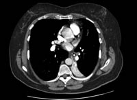
Bronchoscopy
Bronchoscopy determines the exact nature of the foreign body, the location, the degree of airway obstruction, and the associated mucosal abnormalities, such as mucosal oedema and granulation tissue.
Rigid bronchoscopy should be performed in cases of stridor, asphyxia, radio-opaque object on chest radiograph, a history of foreign body aspiration associated with unilaterally decreased breath sounds, localised wheezing, obstructive hyperinflation, or atelectasis.[14]Righini CA, Morel N, Karkas A, et al. What is the diagnostic value of flexible bronchoscopy in the initial investigation of children with suspected foreign body aspiration? Int J Pediatr Otorhinolaryngol. 2007 Sep;71(9):1383-90.
http://www.ncbi.nlm.nih.gov/pubmed/17580093?tool=bestpractice.com
[42]Eber E, Antón-Pacheco JL, de Blic J, et al. ERS statement: interventional bronchoscopy in children. Eur Respir J. 2017 Dec;50(6):1700901.
http://www.ncbi.nlm.nih.gov/pubmed/29242258?tool=bestpractice.com
[43]Chantzaras AP, Panagiotou P, Karageorgos S, et al. A systematic review of using flexible bronchoscopy to remove foreign bodies from paediatric patients. Acta Paediatr. 2022 Jul;111(7):1301-12.
http://www.ncbi.nlm.nih.gov/pubmed/35388522?tool=bestpractice.com
[44]Martinot A, Closset M, Marquette CH, et al. Indications for flexible versus rigid bronchoscopy in children with suspected foreign-body aspiration. Am J Respir Crit Care Med. 1997 May;155(5):1676-9.
http://www.ncbi.nlm.nih.gov/pubmed/9154875?tool=bestpractice.com
In all other circumstances, flexible bronchoscopy should be performed first for diagnostic confirmation.[14]Righini CA, Morel N, Karkas A, et al. What is the diagnostic value of flexible bronchoscopy in the initial investigation of children with suspected foreign body aspiration? Int J Pediatr Otorhinolaryngol. 2007 Sep;71(9):1383-90.
http://www.ncbi.nlm.nih.gov/pubmed/17580093?tool=bestpractice.com
[44]Martinot A, Closset M, Marquette CH, et al. Indications for flexible versus rigid bronchoscopy in children with suspected foreign-body aspiration. Am J Respir Crit Care Med. 1997 May;155(5):1676-9.
http://www.ncbi.nlm.nih.gov/pubmed/9154875?tool=bestpractice.com
In stable children with suspected foreign body aspiration, flexible bronchoscopy has been shown to safely confirm the diagnosis and can be used for therapeutic purposes.[5]Swanson KL, Prakash UB, Midthun DE, et al. Flexible bronchoscopic management of airway foreign bodies in children. Chest. 2002 May;121(5):1695-700.
http://www.ncbi.nlm.nih.gov/pubmed/12006464?tool=bestpractice.com
[43]Chantzaras AP, Panagiotou P, Karageorgos S, et al. A systematic review of using flexible bronchoscopy to remove foreign bodies from paediatric patients. Acta Paediatr. 2022 Jul;111(7):1301-12.
http://www.ncbi.nlm.nih.gov/pubmed/35388522?tool=bestpractice.com
[45]Tang LF, Xu YC, Wang YS, et al. Airway foreign body removal by flexible bronchoscopy: experience with 1027 children during 2000-2008. World J Pediatr. 2009 Aug;5(3):191-5.
http://www.ncbi.nlm.nih.gov/pubmed/19693462?tool=bestpractice.com
[46]Golan-Tripto I, Mezan DW, Tsaregorodtsev S, et al. From rigid to flexible bronchoscopy: a tertiary center experience in removal of inhaled foreign bodies in children. Eur J Pediatr. 2021 May;180(5):1443-50.
http://www.ncbi.nlm.nih.gov/pubmed/33389071?tool=bestpractice.com
Flexible bronchoscopy has been shown to provide rapid and definitive diagnosis of foreign body aspiration in children with persistent wheezing.[47]Cakir E, Ersu RH, Uyan ZS, et al. Flexible bronchoscopy as a valuable tool in the evaluation of persistent wheezing in children. Int J Pediatr Otorhinolaryngol. 2009 Dec;73(12):1666-8.
http://www.ncbi.nlm.nih.gov/pubmed/19733921?tool=bestpractice.com
Rigid bronchoscopy is commonly performed for foreign body removal in children, but it is associated with high rates of negative initial bronchoscopy (11% to 46%).[14]Righini CA, Morel N, Karkas A, et al. What is the diagnostic value of flexible bronchoscopy in the initial investigation of children with suspected foreign body aspiration? Int J Pediatr Otorhinolaryngol. 2007 Sep;71(9):1383-90.
http://www.ncbi.nlm.nih.gov/pubmed/17580093?tool=bestpractice.com
[48]Farrell P. Rigid bronchoscopy for foreign body removal: anaesthesia and ventilation. Pediatr Anaesth. 2004 Jan;14(1):84-9.
http://www.ncbi.nlm.nih.gov/pubmed/14717878?tool=bestpractice.com
Flexible bronchoscopy is a cost-effective procedure in cases of equivocal tracheobronchial foreign body in children, thus avoiding unnecessary rigid bronchoscopy and general anaesthesia.[44]Martinot A, Closset M, Marquette CH, et al. Indications for flexible versus rigid bronchoscopy in children with suspected foreign-body aspiration. Am J Respir Crit Care Med. 1997 May;155(5):1676-9.
http://www.ncbi.nlm.nih.gov/pubmed/9154875?tool=bestpractice.com
[49]Faro A, Wood RE, Schechter MS, et al. Official American Thoracic Society technical standards: flexible airway endoscopy in children. Am J Respir Crit Care Med. 2015 May 1;191(9):1066-80.
https://www.atsjournals.org/doi/full/10.1164/rccm.201503-0474ST
http://www.ncbi.nlm.nih.gov/pubmed/25932763?tool=bestpractice.com
In stable adults, flexible bronchoscopy should be used initially to confirm suspected cases of foreign body aspiration and to attempt removal of the foreign body.[4]Boyd M, Chatterjee A, Chiles C, et al. Tracheobronchial foreign body aspiration in adults. South Med J. 2009 Feb;102(2):171-4.
http://www.ncbi.nlm.nih.gov/pubmed/19139679?tool=bestpractice.com
[50]Ma W, Hu J, Yang M, et al. Application of flexible fiberoptic bronchoscopy in the removal of adult airway foreign bodies. BMC Surg. 2020 Jul 23;20(1):165.
https://bmcsurg.biomedcentral.com/articles/10.1186/s12893-020-00825-5
http://www.ncbi.nlm.nih.gov/pubmed/32703179?tool=bestpractice.com
[51]Sehgal IS, Dhooria S, Ram B, et al. Foreign body inhalation in the adult population: experience of 25,998 bronchoscopies and systematic review of the literature. Respir Care. 2015 Oct;60(10):1438-48.
https://rc.rcjournal.com/content/respcare/60/10/1438.full.pdf
http://www.ncbi.nlm.nih.gov/pubmed/25969517?tool=bestpractice.com
[52]Du Rand IA, Barber PV, Goldring J, et al. British Thoracic Society guideline for advanced diagnostic and therapeutic flexible bronchoscopy in adults. Thorax. 2011 Nov;66(suppl 3):iii1-21.
http://www.ncbi.nlm.nih.gov/pubmed/21987439?tool=bestpractice.com
Flexible bronchoscopy is the method of choice in patients with cervicofacial trauma and in those on mechanical ventilation. In these patients, rigid bronchoscopy should be reserved as a therapeutic approach rather than as a diagnostic tool.