Images and videos
Images


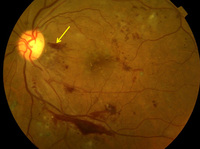


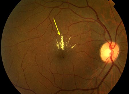
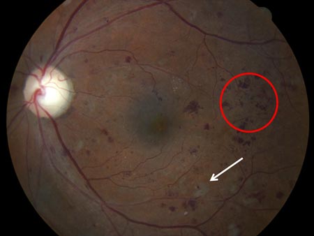
Diabetic retinopathy
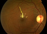
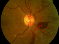
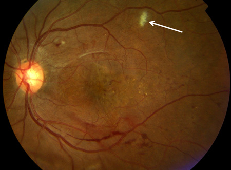
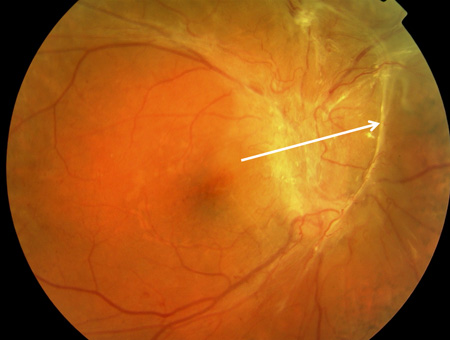
Proliferative diabetic retinopathy: cotton wool spot (white arrow)
Courtesy of Moorfields Photographic Archive; used with permission
See this image in context in the following section/s:

Diabetic retinopathy
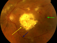
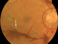
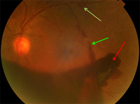
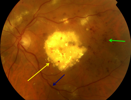
Proliferative diabetic retinopathy: new vessels elsewhere (red arrow), venous beading (blue arrow), intraretinal microvascular abnormality (green arrow)
Courtesy of Moorfields Photographic Archive; used with permission
See this image in context in the following section/s:
Diabetic retinopathy
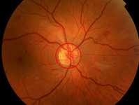
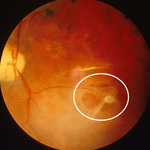
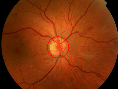
Proliferative diabetic retinopathy: new vessels on the optic disc (red circle)
Courtesy of Moorfields Photographic Archive; used with permission
See this image in context in the following section/s:
Diabetic retinopathy
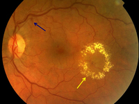
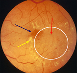
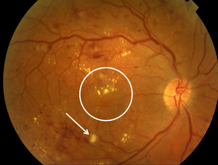
Non-proliferative diabetic retinopathy with macular oedema: exudate (yellow arrow)
Courtesy of Moorfields Photographic Archive; used with permission
See this image in context in the following section/s:

Diabetic retinopathy
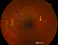
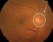
Radiation retinopathy: cotton wool spots (white arrow), optic disc oedema (white circle), venous beading (blue arrow), nerve fibre layer haemorrhage (red arrow)
Courtesy of Moorfields Photographic Archive; used with permission
See this image in context in the following section/s:

Diabetic retinopathy
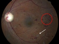
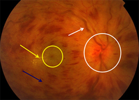
Non-proliferative diabetic retinopathy: intraretinal microvascular abnormality (IRMA; green arrow), venous beading and segmentation (blue arrow), cluster haemorrhage (red circle), featureless retina suggestive of capillary non-perfusion (white ellipse)
Courtesy of Moorfields Photographic Archive; used with permission
See this image in context in the following section/s:

Diabetic retinopathy
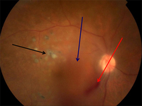
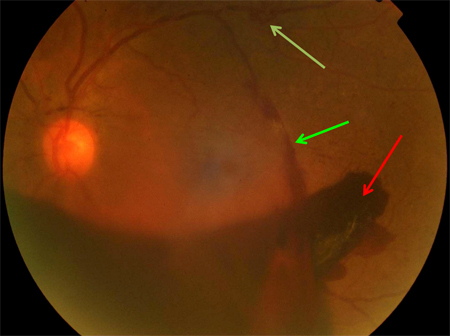
Proliferative diabetic retinopathy: macular laser burns (black arrow), misty vitreous haemorrhage (blue arrow), clot within vitreous haemorrhage (red arrow)
Courtesy of Moorfields Photographic Archive; used with permission
See this image in context in the following section/s:
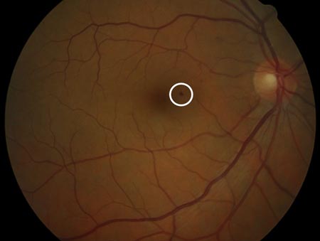
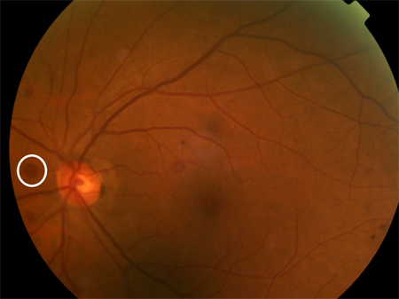
Diabetic retinopathy
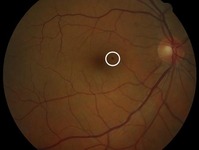
Non-proliferative diabetic retinopathy: blot haemorrhage (white circle)
Courtesy of Moorfields Photographic Archive; used with permission
See this image in context in the following section/s:
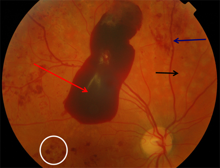
Diabetic retinopathy
Proliferative diabetic retinopathy: retrohyaloid haemorrhage (red arrow), venous beading (blue arrow), cluster haemorrhage (white circle), pan-retinal laser burns (black arrow)
Courtesy of Moorfields Photographic Archive; used with permission
See this image in context in the following section/s:

Diabetic retinopathy
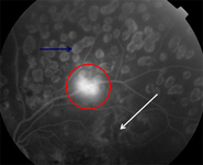
Fluorescein angiography in proliferative diabetic retinopathy: new vessels elsewhere (red circle), capillary non-perfusion (white arrow), pan-retinal laser burns (blue arrow)
Courtesy of Moorfields Photographic Archive; used with permission
See this image in context in the following section/s:
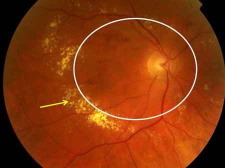
Diabetic retinopathy
Non-proliferative diabetic retinopathy with macular oedema: thickened retina (white ellipse), exudate (yellow arrow)
Courtesy of Moorfields Photographic Archive; used with permission
See this image in context in the following section/s:
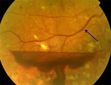
Diabetic retinopathy
Proliferative diabetic retinopathy: traction towards optic disc and consequent total retinal detachment (white block arrow)
Courtesy of Moorfields Photographic Archive; used with permission
See this image in context in the following section/s:

Diabetic retinopathy
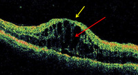
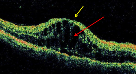
Optical coherence tomography in vitreomacular traction: loss of foveal depression with traction on fovea (in direction of yellow arrow)
Courtesy of Moorfields Photographic Archive; used with permission
See this image in context in the following section/s:
Diabetic retinopathy
Non-proliferative diabetic retinopathy with macular oedema: cotton wool spot (white arrow), thickened retina (white circle)
Courtesy of Moorfields Photographic Archive; used with permission
See this image in context in the following section/s:
Diabetic retinopathy
Proliferative diabetic retinopathy: new vessels elsewhere (white arrow), vitreous (intra-gel) haemorrhage (green arrow), retrohyaloid haemorrhage (red arrow)
Courtesy of Moorfields Photographic Archive; used with permission
See this image in context in the following section/s:

Diabetic retinopathy
Non-proliferative diabetic retinopathy with macular oedema: nerve fibre layer haemorrhage (blue arrow), exudate (yellow arrow)
Courtesy of Moorfields Photographic Archive; used with permission
See this image in context in the following section/s:
Diabetic retinopathy
Non-proliferative diabetic retinopathy with macular oedema: exudate plaque (yellow arrow), cluster haemorrhage (green arrow), venous beading (blue arrow)
Courtesy of Moorfields Photographic Archive; used with permission
See this image in context in the following section/s:

Diabetic retinopathy
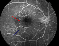
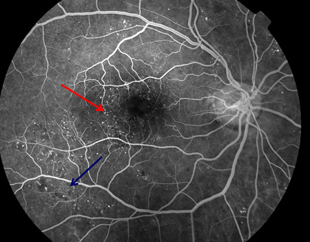
Fluorescein angiogram of non-proliferative diabetic retinopathy: microaneurysms (red arrow), intraretinal microvascular abnormalities (blue arrow)
Courtesy of Moorfields Photographic Archive; used with permission
See this image in context in the following section/s:
Diabetic retinopathy
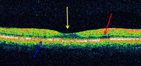
Optical coherence tomography in macular oedema: loss of central foveal depression (yellow arrow), accumulation of fluid within cystoid spaces at fovea (red arrow)
Courtesy of Moorfields Photographic Archive; used with permission
See this image in context in the following section/s:
Diabetic retinopathy
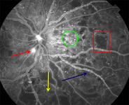
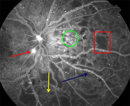
Fluorescein angiogram in proliferative diabetic retinopathy: new vessels on the optic disc (red arrow), capillary non-perfusion (red rectangle), microaneurysms (green circle), venous beading (blue arrow), intraretinal microvascular abnormalities (yellow arrow)
Courtesy of Moorfields Photographic Archive; used with permission
See this image in context in the following section/s:

Diabetic retinopathy
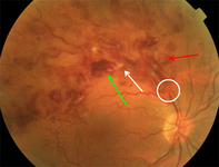
Branch retinal vein occlusion: site of occlusion (white circle), cotton wool spot (white arrow), nerve fibre layer haemorrhage (red arrow), cluster haemorrhage (green arrow)
Courtesy of Moorfields Photographic Archive; used with permission
See this image in context in the following section/s:
Diabetic retinopathy
Fluorescein angiogram of non-proliferative diabetic retinopathy: microaneurysms (red arrow), intraretinal microvascular abnormalities (blue arrow)
Courtesy of Moorfields Photographic Archive; used with permission
See this image in context in the following section/s:
Diabetic retinopathy
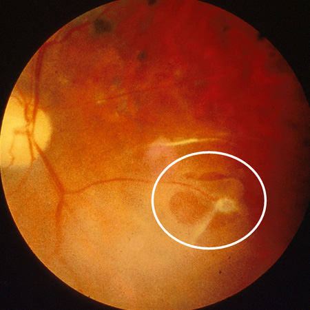
Proliferative diabetic retinopathy: traction tear (white ellipse)
Courtesy of Moorfields Photographic Archive; used with permission
See this image in context in the following section/s:
Diabetic retinopathy
Non-proliferative diabetic retinopathy: cluster haemorrhages (red circle), cotton wool spot (white arrow)
Courtesy of Moorfields Photographic Archive; used with permission
See this image in context in the following section/s:

Diabetic retinopathy
Proliferative diabetic retinopathy: new vessels on the optic disc (red circle), retrohyaloid haemorrhage (red arrow), new vessels elsewhere with fibrosis (white arrow), dot and blot haemorrhage (green arrow)
Courtesy of Moorfields Photographic Archive; used with permission
See this image in context in the following section/s:

Diabetic retinopathy
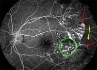
Fluorescein angiography in proliferative diabetic retinopathy. Vascular component of fibrovascular proliferation (red arrows), capillary non-perfusion (yellow arrow), laser burns (green circle)
Courtesy of Moorfields Photographic Archive; used with permission
See this image in context in the following section/s:
Diabetic retinopathy
Proliferative diabetic retinopathy: venous beading (blue arrow)
Courtesy of Moorfields Photographic Archive; used with permission
See this image in context in the following section/s:
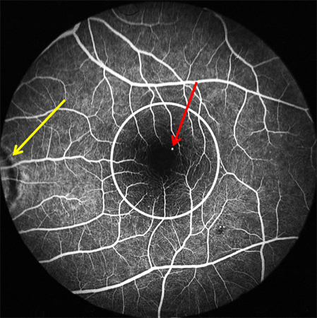
Diabetic retinopathy
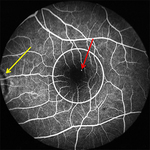
Fluorescein angiogram in mid-venous phase in diabetic retinopathy with microaneurysms only: microaneurysm (red arrow), optic disc (yellow arrow), macula (white circle)
Courtesy of Moorfields Photographic Archive; used with permission
See this image in context in the following section/s:
Diabetic retinopathy
Ocular ischaemic syndrome: scattered posterior-pole and equatorial cluster haemorrhages (white circle)
Courtesy of Moorfields Photographic Archive; used with permission
See this image in context in the following section/s:

Diabetic retinopathy
Non-proliferative diabetic retinopathy with macular oedema: exudate (yellow arrow), microaneurysms (red arrow), thickened retina (white circle), cystic change at macula (blue arrow)
Courtesy of Moorfields Photographic Archive; used with permission
See this image in context in the following section/s:
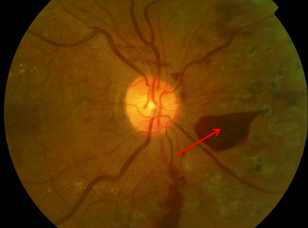
Diabetic retinopathy
Proliferative diabetic retinopathy: retrohyaloid haemorrhage (red arrow)
Courtesy of Moorfields Photographic Archive; used with permission
See this image in context in the following section/s:
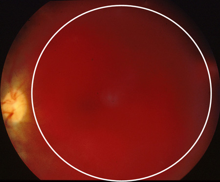
Diabetic retinopathy
Proliferative diabetic retinopathy: extensive vitreous haemorrhage obscuring most of fundus (white circle)
Courtesy of Moorfields Photographic Archive; used with permission
See this image in context in the following section/s:

Diabetic retinopathy
Proliferative diabetic retinopathy: nerve fibre layer haemorrhage (yellow arrow)
Courtesy of Moorfields Photographic Archive; used with permission
See this image in context in the following section/s:
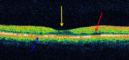
Diabetic retinopathy
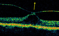
Optical coherence tomogram of normal eye: normal foveal depression at centre of macula (yellow arrow), inner retina (towards centre of eye; red arrow), outer retina (further from centre of eye; blue arrow)
Courtesy of Moorfields Photographic Archive; used with permission
See this image in context in the following section/s:
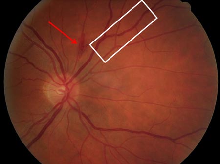
Diabetic retinopathy
Non-proliferative diabetic retinopathy: flame haemorrhage (red arrow), venous beading (white rectangle)
Courtesy of Moorfields Photographic Archive; used with permission
See this image in context in the following section/s:

Diabetic retinopathy
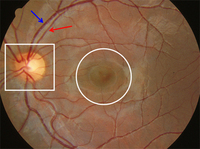
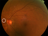
Normal retina left eye: optic disc (white square), macula (white circle), arteriole (red arrow), venule (blue arrow)
Courtesy of Moorfields Photographic Archive; used with permission
See this image in context in the following section/s:

Diabetic retinopathy
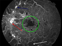
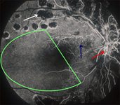
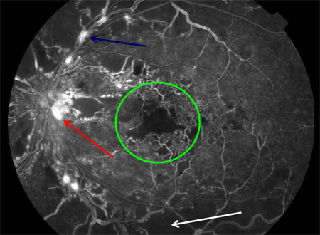
Fluorescein angiogram in proliferative diabetic retinopathy with macular ischaemia: macular ischaemia (green circle), capillary non-perfusion (white arrow), optic disc new vessels (red arrow), venous beading (blue arrow)
Courtesy of Moorfields Photographic Archive; used with permission
See this image in context in the following section/s:
Diabetic retinopathy
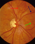
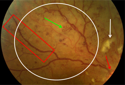
Proliferative diabetic retinopathy: optic disc new vessels (red arrow), intraretinal microvascular abnormality (IRMA; green arrow), cotton wool spot (white arrow), venous beading and segmentation (red rectangle), featureless retina suggestive of capillary non-perfusion (white ellipse)
Courtesy of Moorfields Photographic Archive; used with permission
See this image in context in the following section/s:
Diabetic retinopathy
Fluorescein angiogram in proliferative diabetic retinopathy with macular ischaemia: macular ischaemia (green circle), capillary non-perfusion (white arrow), optic disc new vessels (red arrow), venous beading (blue arrow)
Courtesy of Moorfields Photographic Archive; used with permission
See this image in context in the following section/s:

Diabetic retinopathy
Non-proliferative diabetic retinopathy with macular oedema: exudate (yellow arrow), microaneurysms (red arrow), venous dilatation (blue arrow), cotton wool spot (white arrow)
Courtesy of Moorfields Photographic Archive; used with permission
See this image in context in the following section/s:
Diabetic retinopathy
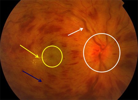
Central retinal vein occlusion: optic disc swelling (white circle), macular oedema (yellow circle), exudate (yellow arrow), nerve fibre layer haemorrhage (blue arrow), venous engorgement and tortuosity (white arrow)
Courtesy of Moorfields Photographic Archive; used with permission
See this image in context in the following section/s:
Use of this content is subject to our disclaimer