Summary
Definition
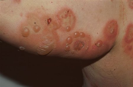
History and exam
Key diagnostic factors
- presence of risk factors
- pruritus
- tense blisters on normal or erythematous skin
Risk factors
- age 60 to 90 years
- major histocompatibility complex (MHC) class II allele (DQB1*0301)
- male sex
Diagnostic investigations
1st investigations to order
- skin biopsy for histopathological evaluation with light microscopy
- skin biopsy for direct immunofluorescence testing
- indirect immunofluorescence test on serum
Emerging tests
- immunoblotting
- immunoprecipitation
- fluorescence overlay antigen mapping (FOAM) technique
Treatment algorithm
Contributors
Authors
Vesna Petronic-Rosic, MD, MSc, MBA
Professor and Chair
Department of Dermatology
Georgetown University
MedStar Washington Hospital Center
Washington
DC
Disclosures
VPR declares that she has no competing interests.
Peer reviewers
Lawrence Parish, MD
Clinical Professor of Dermatology and Cutaneous Biology
Director
Jefferson Center for International Dermatology
Jefferson Medical College
Thomas Jefferson University
Philadelphia
PA
Disclosures
LP declares that he has no competing interests.
Timothy Patton, MD
Assistant Professor of Dermatology
Department of Dermatology
University of Pittsburgh
Pittsburgh
PA
Disclosures
TP declares that he has no competing interests.
Peer reviewer acknowledgements
BMJ Best Practice topics are updated on a rolling basis in line with developments in evidence and guidance. The peer reviewers listed here have reviewed the content at least once during the history of the topic.
Disclosures
Peer reviewer affiliations and disclosures pertain to the time of the review.
References
Key articles
Cotell S, Robinson ND, Chan LS. Autoimmune blistering skin diseases. Am J Emerg Med. 2000;18:288-299. Abstract
Kirtschig G, Middleton P, Bennett C, et al. Interventions for bullous pemphigoid. Cochrane Database Syst Rev. 2010;(10):CD002292.Full text Abstract
Kirtschig G, Khumalo NP. Management of bullous pemphigoid: recommendations for immunomodulatory treatments. Am J Clin Dermatol. 2004;5:319-26. Abstract
Reference articles
A full list of sources referenced in this topic is available here.
Use of this content is subject to our disclaimer