Images and videos
Images


Assessment of coma
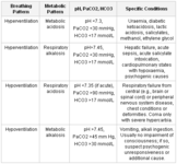
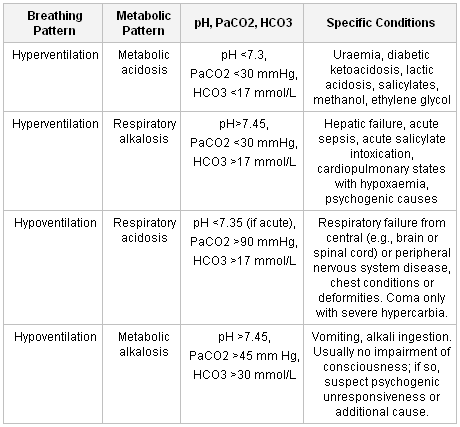
Respiratory abnormalities, blood gas determination, and diagnostic possibilities
Table created by G. Bryan Young, MD; used with permission
See this image in context in the following section/s:

Assessment of coma
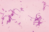
Photomicrograph of Gram-stained Streptococcus species bacteria
From the CDC Public Health Image Library
See this image in context in the following section/s:
Assessment of coma
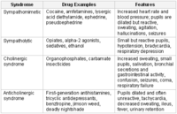
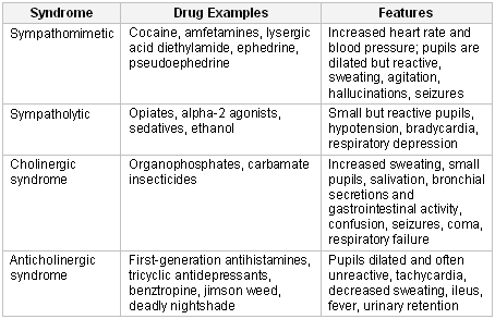
The principal toxidromes, a constellation of features peculiar to certain classes of drugs
Table created by G. Bryan Young, MD; used with permission
See this image in context in the following section/s:

Assessment of coma
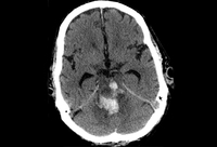
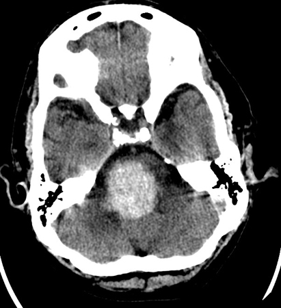
Brainstem haemorrhage in the midbrain that extended from a hypertensive haemorrhage in the pons
From the personal collection of G. Bryan Young, MD; used with permission
See this image in context in the following section/s:
Assessment of coma
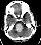
Hypertensive haemorrhage in the pons that ruptured into the fourth ventricle and extended into the midbrain
From the personal collection of G. Bryan Young, MD; used with permission
See this image in context in the following section/s:

Assessment of coma
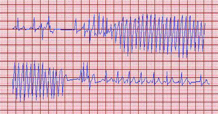
Monomorphic ventricular tachycardia
From the collection of Amar Krishnaswamy; used with permission
See this image in context in the following section/s:
Assessment of coma
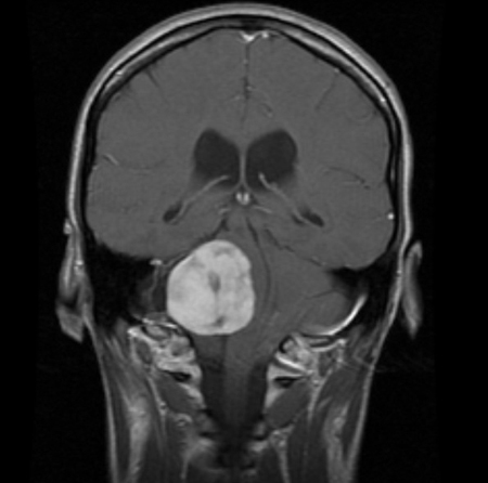
Craniopharyngioma: coronal post-contrast MRI
From the personal collection of Marc C. Chamberlain; used with permission
See this image in context in the following section/s:
Assessment of coma
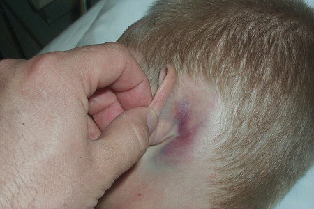
Battle’s sign: superficial ecchymosis over the mastoid process
van Dijk GW. Practical Neurology. 2011;11(1):50-55; used with permission
See this image in context in the following section/s:
Assessment of coma
Torsades de pointes
From the collection of Amar Krishnaswamy; used with permission
See this image in context in the following section/s:
Assessment of coma
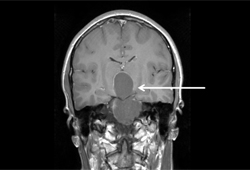
Acoustic neuroma: coronal post-contrast MRI
From the collection of Ryojo Akagami; used with permission
See this image in context in the following section/s:

Assessment of coma
Meningioma: coronal contrast-enhanced image demonstrates meningioma in cavernous sinus on left side
From the personal collection of William T. Couldwell; used with permission
See this image in context in the following section/s:
Assessment of coma
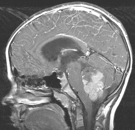
Medulloblastoma: sagittal view MRI showing an avidly enhancing solid and cystic lesion filling the fourth ventricle; obstructive hydrocephalus present
From the collection of Peter B. Storm; used with permission
See this image in context in the following section/s:
Use of this content is subject to our disclaimer