Recommendations
Urgent
This topic covers diagnosis of diabetic ketoacidosis (DKA) in adults.
Consider DKA in:
Any patient with increased thirst, polyuria, recent unexplained weight loss, or excessive tiredness, AND any of the following:[11][20][63][65][66]
Nausea
Vomiting
Abdominal pain
Hyperventilation (Kussmaul respiration)
Dehydration (signs may include dry mucous membranes, tachycardia, decreased skin turgor, slow capillary refill, and hypotension)
Reduced consciousness
Urgently order a venous blood gas, blood ketone (beta-hydroxybutyrate [BOHB]) level, and capillary blood glucose.[2]
These tests should be done at the bedside.
Diagnose DKA if a patient has:[1]
Diabetes - blood glucose ≥11.1 mmol/L OR known diabetes, AND
Ketonaemia - blood ketones ≥3 mmol/L OR ketonuria (2+ or more on standard urine sticks), AND
Acidosis - bicarbonate (HCO3 -) <18 mmol/L AND/OR venous pH <7.3.
Ensure continuous cardiac monitoring and involve senior or critical care support if:[2][20]
There is persistent hypotension (systolic blood pressure <90 mmHg) or oliguria (urine output <0.5 ml/kg/hour) despite intravenous fluids
Stupor and/or coma or Glasgow Coma Scale <12 [ Glasgow Coma Scale Opens in new window ]
Blood ketones (beta-hydroxybutyrate [BOHB]) >6 mmol/L
Venous bicarbonate <10 mmol/L
Venous pH <7.0
Potassium <3.5 mmol/L on admission
Oxygen saturations <92% on air
Pulse >100 bpm or <60 bpm
Anion gap >16 [ Anion Gap Opens in new window ]
The patient is pregnant or has heart or kidney failure or other serious comorbidities.
Involve the specialist diabetes team as soon as possible and definitely within 24 hours.[2]
Key Recommendations
Clinical presentation
Other features of DKA are:
Acetone smell on breath[20]
Smells like pear drops or nail varnish remover
Hypothermia[67]
Most patients with DKA are either normothermic or hypothermic at presentation, even when infection is present, due to peripheral vasodilation.[11] Fever is not a typical feature of DKA, so its presence should prompt consideration of sepsis as a potential precipitating factor. However, bear in mind that sepsis can also present with hypothermia.
History
Ask about causes of DKA. These include:
Discontinuation of insulin (either unintentional or deliberate)[20][64]
Inadequate insulin
New onset of diabetes[20]
Acute illness
Physiological stress
Certain drugs[6][18][20][21][23][26][31][47][48][49][50][70][71][72][73][74]
Corticosteroids
Thiazide diuretics
Sympathomimetics (e.g., dobutamine, terbutaline)
Atypical antipsychotics (e.g., clozapine, olanzapine, risperidone)
Immune checkpoint inhibitors
Cocaine, cannabis, and acute intoxication with alcohol
Sodium-glucose cotransporter-2 (SGLT2) inhibitors (e.g., dapagliflozin, empagliflozin, canagliflozin, ertugliflozin) and the dual SGLT1/SGLT2 inhibitor sotagliflozin.
Examination
Examine the chest:
Auscultate for crepitations or reduced air entry.[75]
These findings may suggest pneumonia - either secondary to aspiration from gastroparesis associated with DKA or as a primary infection triggering DKA - or pulmonary oedema, which is a rare complication of DKA treatment [2][76][77][78] Pulmonary oedema typically occurs several hours after treatment is started and can occur even in patients with normal cardiac function.[2][20]
Assess for signs of dehydration, including:[20]
Dry mucous membranes
Decreased skin turgor or skin wrinkling
Slow capillary refill
Tachycardia with a weak pulse
Hypotension.
Assess conscious level hourly using the Glasgow Coma Scale to monitor for cerebral oedema.[2] [ Glasgow Coma Scale Opens in new window ]
Signs of cerebral oedema include headache, irritability, slowing pulse, rising blood pressure, and reducing conscious level. These may occur several hours after starting treatment.[3][79]
If neurological deterioration occurs, seek immediate critical care input and give mannitol.[80]
Examine the abdomen
Look for an intra-abdominal cause of DKA such as pancreatitis.[42][64]
Be aware that DKA commonly causes abdominal pain and may be mistaken for an acute abdomen.[66][81]
Check the patient’s feet to look for new ulceration or infection.[82]
Check the patient’s skin for rashes, signs of cellulitis, or open wounds that may have precipitated DKA.[83][84]
Investigations
Always order:
Venous blood gas
This will show metabolic acidosis with a raised anion gap. Seek senior or critical care support if pH is <7.0.[2] [ Anion Gap Opens in new window ]
Check the potassium level. Seek senior or critical care support if it is <3.5 mmol/L.[2]
Calculate plasma osmolality. This is typically increased, indicating dehydration.[85]
Blood ketones
Blood glucose
Urea and electrolytes
Full blood count
Consider DKA in:
Any patient with increased thirst, polyuria, recent unexplained weight loss, or excessive tiredness AND any of the following:[11][20][63][65][66]
Nausea
Vomiting
Abdominal pain
Hyperventilation (Kussmaul respiration)
Dehydration (signs may include dry mucous membranes, tachycardia, decreased skin turgor, slow capillary refill, and hypotension)
Reduced consciousness.
This is strongly associated with more severe DKA and a worse prognosis.[89]
Practical tip
DKA is the initial presentation in 6% to 21% of patients with newly diagnosed type 1 diabetes.[1]
DKA is easily missed, especially when it is the initial presentation of diabetes in older patients, or when patients present with other acute medical illnesses such as stroke or myocardial infarction.[79]
Practical tip
While classically associated with type 1 diabetes, DKA can occur in adults with poorly controlled type 2 diabetes, particularly under stressful conditions like infection, acute illness (e.g., myocardial infarction or pancreatitis), or surgery, or in association with drugs such as SGLT2 inhibitors.[1][11]
DKA has also been increasingly documented as a presenting feature of newly diagnosed type 2 diabetes, referred to as ketosis-prone diabetes.[11] Individuals of African, Asian or Hispanic origin and those with obesity or a strong family history of type 2 diabetes appear to be at increased risk.[11][13]
Other features of DKA are:
Acetone smell on breath[20]
The patient’s breath smells like pear drops or nail varnish remover. This is due to high ketone levels.
Bear in mind that a significant proportion of people are unable to smell acetone even if it is present.
Hypothermia
Practical tip
Fever is not a feature of DKA, but DKA may be caused by sepsis. Suspect sepsis as a cause of DKA if there is fever or hypothermia (although hypothermia may also occur in patients with DKA of other aetiologies), hypotension, refractory acidosis, or lactic acidosis.[3]
Ensure continuous cardiac monitoring and involve senior or critical care support if:[2][31]
There is persistent hypotension (systolic blood pressure <90 mmHg) or oliguria (urine output <0.5 ml/kg/hour) despite intravenous fluids
Stupor and/or coma or Glasgow Coma Scale <12 [ Glasgow Coma Scale Opens in new window ]
Blood ketones (beta-hydroxybutyrate [BOHB]) >6 mmol/L
Venous bicarbonate <10 mmol/L
Venous pH <7.0
Potassium <3.5 mmol/L on admission
Oxygen saturations <92% on air
Pulse >100 bpm or <60 bpm
Anion gap >16 [ Anion Gap Opens in new window ]
The patient is pregnant or has heart or kidney failure or other serious comorbidities.
Evidence: Clinical predictors of outcomes in DKA
Prognostic factors for survival in patients with DKA are unclear.
There is limited evidence from a case series of patients with DKA in India for several favourable prognostic indicators, including being male, having lower APACHE scores, and having lower serum phosphate levels on presentation.
A case series assessed 270 patients hospitalised with DKA in India over 2 years.[92]
It found that survival was more likely among males than females (odds ratio [OR] 7.93, 95% CI 3.99 to 13.51).[92]
Other favourable prognostic factors in multivariate analysis (adjusting for type of diabetes, blood pressure, total leukocyte count, urea, serum creatinine, serum magnesium, serum osmolality, serum glutamic oxaloacetic transaminases, serum glutamic pyruvic transaminases, and serum albumin) were lower APACHE scores (OR 2.86, 95% CI 1.72 to 7.03) and lower serum phosphate (OR 2.71, 95% CI 1.51 to 6.99) at presentation.[92]
However, this study reported a high overall mortality rate and may not be representative of UK or European populations.[92]
Involve the specialist diabetes team as soon as possible and definitely within 24 hours.[2]
The specialist diabetes team should also be involved in the assessment of the cause of DKA.
It is unsafe to manage DKA without the specialist diabetes team and could compromise patient care.[2]
Ask about possible causes of DKA. These include:
Discontinuation of insulin (unintentional or deliberate; second most common cause of DKA)[20][64]
Ask sensitively about reasons for deliberate discontinuation of insulin, which may include fear of weight gain or hypoglycaemia, financial barriers, and psychological factors such as needle phobia and stress.[20][63]
Younger patients with type 1 diabetes may omit insulin due to fear of hypoglycaemia, weight gain, eating disorders, or the stress of having a chronic disease. These factors may account for 20% of episodes of recurrent DKA.[93]
Inadequate insulin
New onset of diabetes[20]
Acute illness
Physiological stress
Past medical history
History of diabetes
DKA is most common in people with type 1 diabetes but can occur in those with type 2 diabetes.[11]
Drug history[20]
Drugs that may cause DKA include:[6][18][20][21][23][26][31][47][48][49][50][70][71][72][73][74]
Corticosteroids (increase insulin resistance)
Thiazide diuretics (unclear cause but may increase insulin resistance, inhibit glucose uptake, and decrease insulin release)
Sympathomimetics, such as dobutamine or terbutaline (alter glucose metabolism)
Atypical antipsychotics, such as clozapine, olanzapine, and risperidone (alter glucose metabolism)
Immune checkpoint inhibitors (cause insulin deficiency)
Cocaine, cannabis, and acute intoxication with alcohol
SGLT2 inhibitors (e.g., dapagliflozin, empagliflozin, canagliflozin, ertugliflozin) and the dual SGLT1/SGLT2 inhibitor sotagliflozin (prevent reabsorption of glucose and facilitate its excretion in urine).
Practical tip
Diagnosis of DKA in pregnancy is often delayed because it can occur at lower blood glucose levels (including euglycaemic DKA) and faster than in non-pregnant patients.[2][98]
DKA in pregnancy may present with abdominal pain; always consider as a possible alternative to pre-term or term labour.[2]
DKA usually occurs in the second and third trimesters due to increased insulin resistance.[98] Pregnant women suspected of having DKA should receive care from both the obstetric and medical (or diabetes) teams.[2]
Examine the chest.
Look for hyperventilation (Kussmaul respiration).[11][20]
This is a late sign of DKA and occurs with more severe acidosis.
Characterised by deep sighing respirations at a slow or normal rate.
Auscultate for crepitations or reduced air entry.[75]
This may be due to pneumonia, which can be caused by aspiration from gastroparesis in DKA or a primary infection.[76][77][78]
Basal crepitations are also a sign of pulmonary oedema or acute respiratory distress syndrome (ARDS) secondary to fluid overload. This is an uncommon complication of treatment for DKA.[2]
Check for signs of dehydration. These include:[20]
Dry mucous membranes
Decreased skin turgor or skin wrinkling
Slow capillary refill
Tachycardia with a weak pulse
Hypotension.
Assess conscious level hourly using the Glasgow Coma Scale to monitor for cerebral oedema.[2] [ Glasgow Coma Scale Opens in new window ]
Mental status can range from alert in mild DKA to coma in severe DKA.[67]
Cerebral oedema can develop during treatment of DKA due to rapid correction of hyperglycaemia.[2]
Signs include headache, irritability, slowing pulse, rising blood pressure, and reducing conscious level. These may occur several hours after starting treatment.[3][79]
Papilloedema is a late sign of cerebral oedema.[3]
If neurological deterioration occurs, seek urgent input from the critical care team and give mannitol without delay.[80]
Cerebral oedema has a mortality rate of 70%. It is most common in children and adolescents but can occur rarely in adults.[63]
Examine the abdomen for a possible cause of DKA, such as pancreatitis.[42][64] DKA can both cause and mimic an acute abdomen.[66][81] DKA must be excluded prior to any emergency surgery.
Look for abdominal distension, which may indicate bowel obstruction.[99]
Palpate the abdomen to check for rebound tenderness and guarding caused by irritation of the peritoneum.[99]
Auscultate for bowel sounds.[100]
Hyperactive ‘tinkling’ bowel sounds may be present in early bowel obstruction.
Reduced or absent bowel sounds may be present in late bowel obstruction, perforated viscus, haemoperitoneum, or any cause of peritoneal inflammation.
Perform a rectal examination.[99]
Ensure you take a chaperone with you.
Assess for occult or frank blood, pain, or a mass.
Practical tip
The severity of abdominal pain caused directly by DKA correlates strongly with the severity of the metabolic acidosis.[66]
Check the patient’s feet to look for new ulceration or infection.[82]
Practical tip
Check the feet for loss of protective sensation in any patient with diabetes.
Follow your local guidelines, but a quick simple test is the Ipswich Touch Test©️, which involves lightly touching/resting the tip of the index finger for 1 to 2 seconds on the tips of the first, third, and fifth toes and the dorsum of the hallux.[101]
If your patient has reduced sensation, they are at high risk of pressure ulceration. Inform the nursing staff and provide pressure-relieving devices.
A daily heel check for signs of pressure trauma should be done by nursing or healthcare assistant staff.
There is a debate about whether compression stockings should or should not be used in people with diabetes - do not use them if there is vascular disease.
Check the patient’s skin for rashes and signs of cellulitis or open wounds.
Diagnose DKA if a patient has:[1]
Diabetes - blood glucose ≥11.1 mmol/L OR known diabetes, AND
Ketonaemia - blood ketones (beta-hydroxybutyrate [BOHB]) ≥3 mmol/L OR ketonuria (2+ or more on standard urine sticks), AND
Acidosis - bicarbonate (HCO3 -) <18 mmol/L AND/OR venous pH <7.3.
Practical tip
Assessment of glucose, ketones, and electrolytes, including bicarbonate and venous pH, should be done at or near the bedside.[2]
Order laboratory measurements in certain circumstances, such as when blood glucose or ketone meters are ‘out of range’.
Practical tip
Rarely, patients present with euglycaemic DKA (EDKA) and have a normal blood glucose level.[20][103] Always use pH and ketones to guide diagnosis and management in patients with known diabetes (rather than relying solely on a ‘glucose-centric’ approach).[2]
Exclude other causes of an anion gap metabolic acidosis before confirming EDKA.
The mechanism of EDKA is unclear but may be due to decreased insulin secretion with increased counter-regulatory hormone secretion (cortisol, glucagon, catecholamines, and growth hormone).[24]
Possible precipitants of EDKA are pregnancy, starvation, alcohol use, insulin pumps, and SGLT2 or SGLT1/SGLT2 inhibitors.[24][103]
Patients with EDKA secondary to treatment with an SGLT2 or SGLT1/SGLT2 inhibitor may have less polyuria and polydipsia due to a lower glucose level. They may instead present with malaise, anorexia, tachycardia, or tachypnoea, with or without fever.[20]
Always order the following investigations
Venous blood gas[2]
This will show metabolic acidosis with a raised anion gap. [ Anion Gap Opens in new window ]
Anion gap >16 indicates severe DKA.
Use the pH to determine the severity of DKA.
pH ≥7.0 indicates mild or moderate DKA.
pH <7.0 indicates severe DKA. Discuss these patients with critical care.
Use the potassium level on venous blood gas to replace potassium if ≤5.0 mmol/L.[1] Discuss with a senior or critical care if potassium is <3.5 mmol/L.[2]
Practical tip
Exercise caution when interpreting a potassium result obtained from a blood gas analyser.
Blood gas analysers are often used as point-of-care tests, providing rapid results (e.g., within minutes) for potassium levels, which is crucial in the initial management of DKA to guide insulin infusion.
Studies have shown that measurements from blood gas analysis may underestimate serum potassium levels in DKA.[104][105]
Be aware of these potential limitations and interpret results in conjunction with clinical judgement and other laboratory data, especially when making decisions about potassium replacement therapy.
Calculate plasma osmolality.
Plasma osmolality is typically increased in DKA and is an indication of dehydration.[85]
Evidence: Use of a venous versus arterial blood gas
Venous blood gas measurements are widely used instead of arterial blood gas measurements and evidence from case studies suggests there is sufficient agreement between them, when combined with other clinical findings, to use a venous blood gas to guide initial treatment.
A clinical review article aimed to answer the question “can venous blood gas analysis replace arterial blood gas analysis in emergency care?[106]
Venous blood gas testing may have a lower risk of serious adverse events (e.g., vascular occlusion or infection), is less painful for the patient, and is technically easier to perform than arterial blood gas testing.
There is little difference in pH values between venous and arterial samples (based on 13 studies; 2009 participants, with 3 studies [295 patients] in patients with DKA).[107]
Bicarbonate values also show close agreement between venous and arterial samples (8 studies; 1211 patients).[107]
Agreement for PCO 2 is poor and unpredictable (8 studies; 965 patients), but a venous PCO 2 ≤45 mmHg (6 kPa) reliably excludes clinically significant hypercarbia (4 studies; 529 patients; 100% sensitivity).[107]
Agreement on lactate is close enough to categorise as high or normal (3 studies; 338 patients).[108]
Evidence regarding arteriovenous agreement for base excess is unclear (2 studies; 429 patients; only 1 study reporting close agreement).[109][110]
If data from the venous blood gas does not appear to match the patient’s clinical condition, an arterial blood gas should be performed.[106]
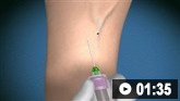
How to take a venous blood sample from the antecubital fossa using a vacuum needle.
Blood ketones[2]
This will show ketonaemia (ketones ≥3 mmol/L) in DKA.[2]
Use urinary ketones if near patient blood ketone testing is unavailable. This will show ketonuria (2+ or more on standard urine sticks).[2]
Practical tip
Bear in mind that a patient’s medications can cause errors in detecting ketone bodies.
Some drugs, such as the ACE inhibitor captopril, contain sulfhydryl groups that can react with the reagent in the nitroprusside test (used to test for ketone bodies) to give a false-positive reaction.[112] Therefore, use clinical judgement and other biochemical tests in patients who are taking these medications.
Blood glucose
Hyperglycaemia (blood glucose ≥11.1 mmol/L) is common.[1]
Be aware that some patients can present with euglycaemic DKA and have a normal blood glucose.[86]
Always use pH and ketones alongside glucose to guide diagnosis and management.
Manage euglycaemic DKA in the same way as hyperglycaemic DKA.[2]
Urea and electrolytes
Hyponatraemia is common in DKA.[2]
Hypernatraemia with hyperglycaemia indicates severe dehydration.[88]
Hyperkalaemia is common but hypokalaemia is an indicator of severe DKA.[2]
Hypokalaemia on arrival indicates severe total-body potassium deficit and is an indicator of severe DKA.[88] This is because the total body potassium concentration is low due to increased diuresis.
Hyperkalaemia is due to an extracellular shift of potassium caused by insulin insufficiency, hypertonicity, and acidosis.[88]
Hypomagnesaemia and hypophosphataemia may also be present.[2][87]
Full blood count
Leukocytosis is common in DKA and correlates with blood ketone levels.[88]
However, leukocytosis more than 25 × 10⁹/L (25,000/microlitre) may indicate infection and requires further investigation.[88]
Consider ordering the following investigations
Urinalysis
Order if near-patient testing for blood ketones is unavailable or you suspect a urinary tract infection.[2]
Shows ketonuria (2+ or more on standard urine sticks) in patients with DKA.[2]
May be positive for glucose.[113]
Other findings include leukocytes and nitrites in the presence of infection, and myoglobinuria and/or haemoglobinuria in rhabdomyolysis (which is present in about 10% of patients with DKA).[113][114][115]
ECG
Use to look for cardiac precipitants of DKA such as myocardial infarction.[2]
Findings may include abnormal T or Q waves or ST segment changes.[116]
Look for cardiac effects of electrolyte abnormalities.

How to record an ECG. Demonstrates placement of chest and limb electrodes.
Pregnancy test
Order in all women of childbearing age.[2]
Amylase and lipase
Non-specific elevations of amylase can be seen in DKA.[119]
In one study, amylase was elevated in 21% of patients with DKA.[42]
Serum lipase is usually normal.
Cardiac enzymes
Order troponin T or I if you suspect myocardial infarction as a precipitant.[120]
Creatinine kinase
Elevated if rhabdomyolysis is present. This is present in around 10% of patients with DKA.[114]
Chest x-ray
Order if there are reduced oxygen saturations.[2]
Signs of pulmonary oedema include pleural effusions, interstitial and alveolar oedema, prominent superior vena cava, Kerley B lines, and dilated upper lobe blood vessels.[121]
Consolidation occurs in pneumonia.
Liver function tests (LFTs)
Useful to screen for an underlying hepatic precipitant of DKA. Abnormal LFTs may indicate underlying liver disease (e.g., metabolic dysfunction-associated steatotic liver disease or congestive heart failure).[122][123] Chronic liver disease is a risk factor for euglycaemic DKA.[124]
Blood, urine, and sputum cultures
How to take a venous blood sample from the antecubital fossa using a vacuum needle.
How to record an ECG. Demonstrates placement of chest and limb electrodes.
Use of this content is subject to our disclaimer