Images and videos
Images


Assessment of back pain
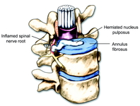
Herniated nucleus pulposus causing nerve root impingement. Radicular symptoms may result from chemical mediators released by degenerative discs or mechanical compression of the nerve root
BMJ 2008;337:a2718; used with permission
See this image in context in the following section/s:

Assessment of back pain
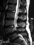
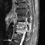
Magnetic resonance imaging of spinal stenosis: arrow points to the moderately stenotic spinal canal caused by hypertrophic facets and ligament flavum
Courtesy of Dr K. Singh; used with permission
See this image in context in the following section/s:
Assessment of back pain
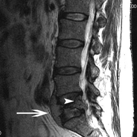
Magnetic resonance imaging of lymphoma: arrowhead indicates a soft-tissue mass protruding into the spinal canal. Arrow points to the tumour protruding anterior to the L5 vertebral body
Courtesy of Dr K. Singh; used with permission
See this image in context in the following section/s:

Assessment of back pain
Magnetic resonance imaging of spinal stenosis: (A) demarcates the normal sagittal diameter of the spinal canal. (B) demarcates severe narrowing of the spinal canal
Courtesy of Dr K. Singh; used with permission
See this image in context in the following section/s:

Assessment of back pain
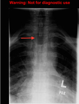
The 'winking owl' sign (arrow): asymmetrical appearance of spine on plain x-rays caused by destruction of the pedicle
Created by BMJ Publishing Group
See this image in context in the following section/s:

Assessment of back pain
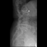
X-ray of a compression fracture: a lateral x-ray of an L2 compression fracture (A). Wedging of the vertebral body is seen
Courtesy of Dr K. Singh; used with permission
See this image in context in the following section/s:

Assessment of back pain
X-ray of tumour: lymphoma (A) destroying the L5 vertebra
Courtesy of Dr K. Singh; used with permission
See this image in context in the following section/s:

Assessment of back pain
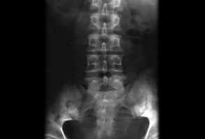
Spine x-ray: the 'winking owl' sign (asymmetrical appearance caused by destruction of the pedicle)
Courtesy of Dr D. Park; used with permission
See this image in context in the following section/s:
Assessment of back pain
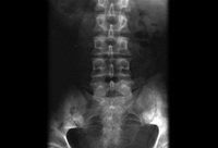
Plain x-ray showing bilateral sacroiliitis in a patient with ankylosing spondylitis
BMJ 2006;333;581-585. © BMJ Publishing Group Ltd 2009
See this image in context in the following section/s:
Assessment of back pain
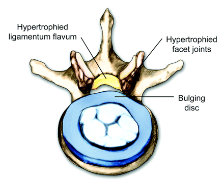
Axial view of a vertebral body showing central spinal stenosis
BMJ 2008;337:a2718; used with permission
See this image in context in the following section/s:

Assessment of back pain
Magnetic resonance imaging of osteomyelitis: T11-T12 disc space is involved with discitis (A). There is bony involvement of both vertebrae indicated by high T2 signal of the vertebral bodies. Arrow indicates a normal healthy vertebral disc
Courtesy of Dr K. Singh; used with permission
See this image in context in the following section/s:
Assessment of back pain
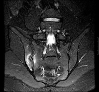
Coronal STIR (short tau inversion recovery) magnetic resonance image showing unilateral (right) sacroiliitis
BMJ 2006;333;581-585. © BMJ Publishing Group Ltd 2009
See this image in context in the following section/s:
Assessment of back pain
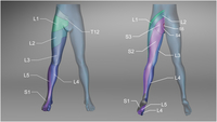
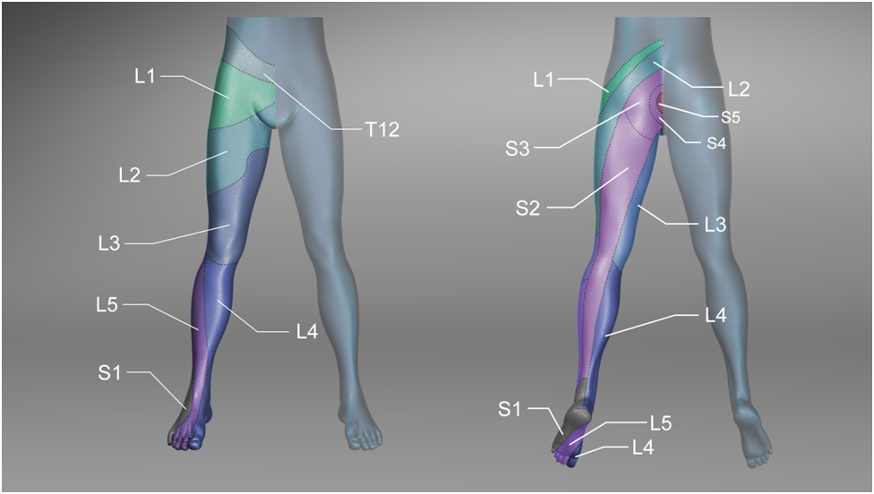
Sensory dermatomes of the lower back and leg
Created by BMJ Knowledge Centre
See this image in context in the following section/s:
Videos
 Inspection of the back
How to perform an inspection examination of the back, including inspection of gait and posture
 Physical examination of the back demonstration
A GP demonstrates how to perform a physical examination of the back
 Neurological examination of the back demonstration
A GP shows how to perform a neurological examination of the back
Use of this content is subject to our disclaimer