Aetiology
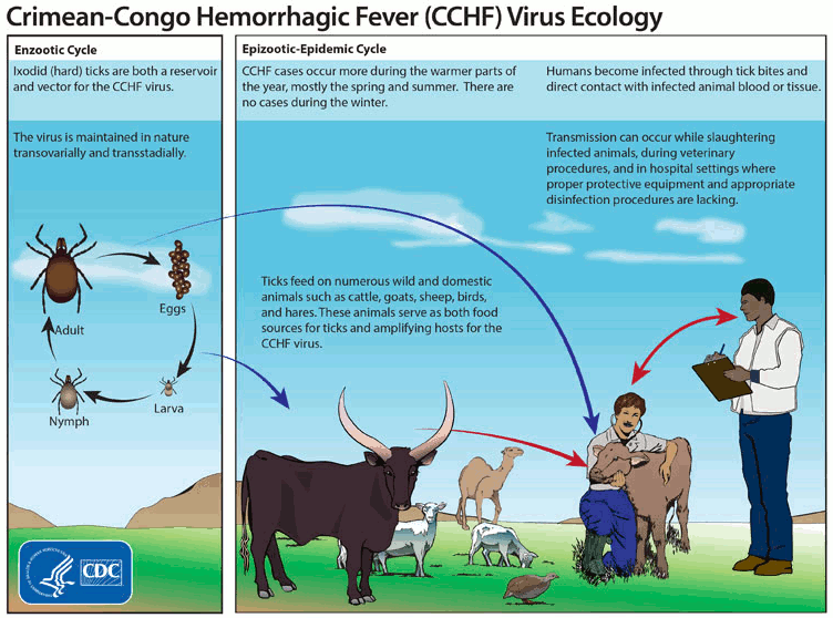
CCHF virus is a single-stranded RNA virus of the Nairoviridae family (genus Orthonairovirus), and is related to the hantavirus and Rift Valley fever virus. CCHF virus circulates in an enzootic tick-vertebrate-tick cycle and there is evidence that the virus causes disease in animals. Humans become infected through the bites of ticks, by contact with a patient with CCHF during the acute phase of infection, or by contact with blood or tissues from viraemic livestock.[2][32]
Ticks of the family Ixodidae (genus Hyalomma) are considered to be the main vectors that spread the virus to humans. The occurrence of CCHF closely approximates the known world distribution of Hyalomma ticks, with phylogenetic analyses revealing relations between strains from distant outbreaks. Although ticks of the genus Hyalomma are recognised as the main vector/reservoir of the virus, it has also been detected in ticks of the genuses Ornithodoros and Amblyomma. The role of tick species other than Hyalomma is not fully understood and requires further investigation.[33]
In endemic areas, antibody surveys in livestock have confirmed high prevalence among cattle and sheep. Smaller wildlife species such as hedgehogs and hares, which act as hosts for the tick vectors’ immature stages, more commonly demonstrate CCHF viral infection.[1] Birds may play a role in transporting CCHF virus-infected ticks between countries.[34] Entry of Hyalomma ticks to Great Britain was assessed after the outbreak in Spain in 2016.[35] A Hyalomma tick was detected on a horse for the first time in the UK in 2018; the risk of CCHF in the UK is still considered very low.[36]
Human-to-human transmission occurs via contact with body fluids from infected patients. Healthcare workers and household contacts are at risk of infection if they are exposed to the body fluids of an infected patient without appropriate protective equipment.
Multiple CCHF virus strains have been reported.[37][38] All except one, AP92, cause symptomatic disease.
[Figure caption and citation for the preceding image starts]: Crimean-Congo haemorrhagic fever virus ecologyCenters for Disease Control and Prevention (CDC) [Citation ends].
Pathophysiology
The incubation period for CCHF virus ranges from 1 to 9 days, but 30 days of incubation period was reported from Turkey.[39] Following inoculation, the CCHF virus initially replicates in local tissues, including dendritic cells, before migrating to regional lymph nodes and disseminating through monocytes to tissues and organs including the liver, spleen, and lymph nodes. Migration of tissue macrophages leads to secondary infection of permissive parenchymal cells. Lymphocytes remain free of infection, but, over the course of the illness, may be destroyed in large numbers through apoptosis as occurs in other forms of septic shock.[40][41]
Microvascular instability and impaired haemostasis are the hallmarks of CCHF virus infection. Impaired haemostasis may be the result of platelet, endothelial cell, and/or coagulation factor dysfunction. Reduction in coagulation factors may be due to hepatic dysfunction and/or disseminated intravascular coagulopathy, which is frequently noted in CCHF virus infections. In addition, haemorrhagic diathesis may occur through direct damage of endothelial cells and platelets, and/or indirectly through immunological and inflammatory pathways.[42][43][44] These changes seem to be due to the release of pro-inflammatory mediators such as chemokines and cytokines from virus-infected macrophages and monocytes.[45][46] Tissue damage may be the result of direct necrosis of infected cells or, indirectly, through apoptosis of immune cells. Hepatocytes are particularly affected in CCHF virus infection.[40][47]
CCHF viral load has consistently been shown to correlate with patient outcomes. A viral load >10⁸ copies/mL has been shown to be strongly associated with severe disease and death.[48][49][50]
Use of this content is subject to our disclaimer