History and exam
Key diagnostic factors
common
presence of risk factors
Risk factors for abdominal wall defects include smoking and maternal infection during pregnancy.
maternal age <20 years (gastroschisis)
maternal age >35 years (omphalocele)
elevated maternal serum alpha-fetoprotein (gastroschisis)
positive antenatal ultrasound
Abdominal wall defects of the fetus are usually detected on routine antenatal ultrasound in the second trimester, when an abdominal mass may be visualised outside of the abdominal wall.
During second-trimester ultrasonography, these defects are characterised by abdominal wall masses and echogenic bowel outside of the abdominal wall. In gastroschisis, dilated, fluid-filled loops of bowel floating freely in the amniotic fluid, and, in some cases, intestinal atresia, may be detected.
fetal chromosomal abnormalities (omphalocele)
If evidence of omphalocele is confirmed on ultrasonography, invasive procedures, such as amniocentesis performed at 15 to 20 weeks' gestation or chorionic villus sampling performed at 10 to 12 weeks' gestation, may be undertaken to evaluate the possibility of associated chromosomal abnormalities such as trisomy 13, trisomy 18, trisomy 21, Turner syndrome, Klinefelter syndrome, or triploidy.[9][17][20]
abdominal contents external to the abdominal wall
Omphaloceles are abdominal wall defects ranging from 4 to 12 cm in size and can be located centrally, or in the epigastric or hypogastric regions. In omphalocele, as the abdominal contents have a protective membranous covering in utero, the intestines are usually healthy at birth.
In gastroschisis, the lack of a protective membranous covering causes the abdominal contents to be free-floating in utero, leading to a chemical reaction that creates a thick inflammatory film or peel overlying the intestine. Gastroschisis is commonly associated with intestinal atresia, which occurs in 10% to 15% of cases, and is related to ischaemia of the exposed gut caused by constriction of its mesenteric blood supply at the level of the abdominal wall defect.[31][Figure caption and citation for the preceding image starts]: Note the membrane covering the abdominal contents in this omphaloceleFrom collection of J.J. Tepas III, MD, FACS, FAAP [Citation ends].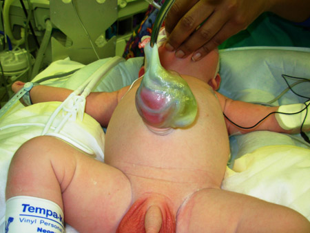 [Figure caption and citation for the preceding image starts]: Extruded gut in abdominal wall defectFrom collection of J.J. Tepas III, MD, FACS, FAAP [Citation ends].
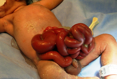
Risk factors
strong
maternal age <20 years (gastroschisis)
male sex of neonate (gastroschisis)
maternal age >35 years (omphalocele)
smoking
Evidence supporting an association between smoking during pregnancy and the development of abdominal wall defects in the fetus is well described for both gastroschisis and omphalocele. It is suggested that cigarette smoke causes vasoconstriction that contributes to placental insufficiency and failed development of the vascular system.[11][13][14][15] The incidence of gastroschisis in babies of teenage mothers who are smokers is increasing.[3][4][9][10]
weak
maternal infection during pregnancy
Maternal infections and illness during pregnancy are seen in mothers who deliver infants with omphalocele and gastroschisis. It is proposed that maternal illness can contribute to placental insufficiency that may, in turn, contribute to the development of abdominal wall defects.[4] Genitourinary infection during pregnancy has also been shown to be associated with abdominal wall defects.[16]
Use of this content is subject to our disclaimer