Investigations
1st investigations to order
antinuclear antibodies, double-stranded (ds) DNA
Test
Should be performed in all patients presenting with DLE lesions to detect progression to systemic lupus erythematosus (SLE) as early as possible.
Antinuclear antibody (ANA) titre is low in 30% to 40% of patients.
High-titre ANA, anti-dsDNA antibodies, and anti-ribonucleoprotein antibodies may suggest SLE or overlapping connective tissue disease. Anti-Ro (SS-A) antibodies can occasionally be found in patients with DLE, whereas anti-La (SS-B) antibodies are distinctly unusual in patients with isolated DLE.
Result
positive or negative
FBC
Test
Should be performed in all patients presenting with DLE lesions.
Although typically normal, a few patients may have mild leukopenia. Modest to severe leukopenia may suggest underlying systemic lupus erythematosus.
Result
normal or mild leukopenia
ESR
Test
Should be performed in all patients presenting with DLE lesions.
Moderate or marked elevation is more likely to be associated with active systemic lupus erythematosus or underlying infection.
Result
normal or slightly elevated
urea and electrolytes
Test
Should be performed in all patients presenting with DLE lesions to exclude any systemic involvement, particularly renal impairment, associated with systemic lupus erythematosus.
Result
normal
urinalysis
Test
Should be performed in all patients presenting with DLE lesions to exclude any systemic involvement, particularly renal impairment, associated with systemic lupus erythematosus.
Result
normal
Investigations to consider
skin biopsy
Test
Confirms diagnosis. Usually indicated if the diagnosis is in doubt. [Figure caption and citation for the preceding image starts]: Skin biopsy showing hyperkeratosis, thinning of the epidermis, degeneration of the basal layer of the epidermis (arrow), and a predominantly lymphocytic inflammatory cell infiltrate in the dermisFrom the collection of Dr K. Blessing, used with permission [Citation ends].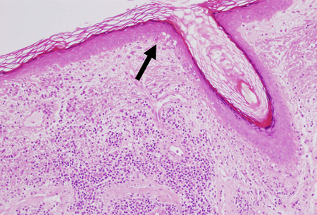
Result
prominent hyperkeratosis, follicular plugging, vacuolar degeneration, thickened epidermal basement membrane, peri-appendageal inflammation
Use of this content is subject to our disclaimer