Images and videos
Images


Assessment of secondary amenorrhoea
World Health Organization classification of amenorrhoea
Created by BMJ Knowledge Centre
See this image in context in the following section/s:

Assessment of secondary amenorrhoea
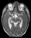
T2-weighted axial MRI scan showing a lesion in the pituitary fossa (arrow), displaying heterogeneous signal intensity suggesting recent apoplexy
BMJ Case Reports 2009; doi:10.1136/bcr.09.2008.0902
See this image in context in the following section/s:
Assessment of secondary amenorrhoea
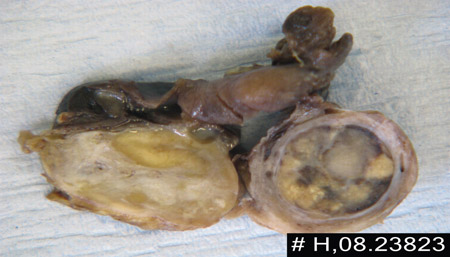
Androgen-secreting tumour in cut section of right ovary
BMJ Case Reports 2009; doi:10.1136/bcr.11.2008.1286
See this image in context in the following section/s:
Assessment of secondary amenorrhoea
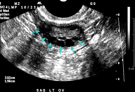
Polycystic ovarian ultrasound
From the collection of Dr M.O. Goodarzi; used with permission
See this image in context in the following section/s:
Assessment of secondary amenorrhoea
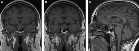
(A) Coronal T1-weighted MRI scan showing a pituitary mass with expansion of the pituitary fossa (B) Coronal T1-weighted MRI scan showing a pituitary mass extending into the cavernous sinus, particularly on the right (C) Sagittal T1-weighted MRI scan of the pituitary tumour
BMJ Case Reports 2009; doi:10.1136/bcr.08.2009.2193
See this image in context in the following section/s:
Assessment of secondary amenorrhoea
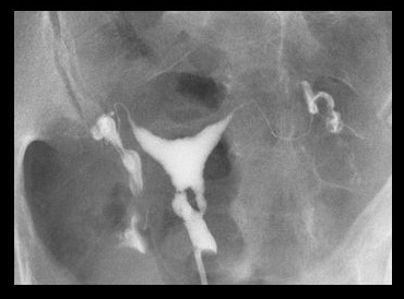
Lower uterine segment in patient with Asherman's syndrome, seen on hysterosalpingogram
From the collection of Dr Meir Jonathon Solnik
See this image in context in the following section/s:
Use of this content is subject to our disclaimer