A investigação diagnóstica da leucemia de células pilosas (LCP) inclui uma história completa e exame físico, exames laboratoriais (incluindo exame de sangue periférico para identificar células leucêmicas [pilosas]), avaliação da medula óssea (incluindo análise morfológica, imunofenotípica e molecular) e, em alguns casos, exames de imagem.[7]Parry-Jones N, Joshi A, Forconi F, et al. Guideline for diagnosis and management of hairy cell leukaemia (HCL) and hairy cell variant (HCL-V). Br J Haematol. 2020 Dec;191(5):730-7.
https://www.doi.org/10.1111/bjh.17055
http://www.ncbi.nlm.nih.gov/pubmed/33053222?tool=bestpractice.com
[33]Grever MR, Abdel-Wahab O, Andritsos LA, et al. Consensus guidelines for the diagnosis and management of patients with classic hairy cell leukemia. Blood. 2017 Feb 2;129(5):553-60.
https://www.doi.org/10.1182/blood-2016-01-689422
http://www.ncbi.nlm.nih.gov/pubmed/27903528?tool=bestpractice.com
[34]National Comprehensive Cancer Network. NCCN clinical practice guidelines in oncology: hairy cell leukemia [internet publication].
https://www.nccn.org/professionals/physician_gls/default.aspx
Quadro clínico
Os pacientes podem apresentar fadiga, letargia, infecção e/ou perda de peso.[7]Parry-Jones N, Joshi A, Forconi F, et al. Guideline for diagnosis and management of hairy cell leukaemia (HCL) and hairy cell variant (HCL-V). Br J Haematol. 2020 Dec;191(5):730-7.
https://www.doi.org/10.1111/bjh.17055
http://www.ncbi.nlm.nih.gov/pubmed/33053222?tool=bestpractice.com
[35]Quest GR, Johnston JB. Clinical features and diagnosis of hairy cell leukemia. Best Pract Res Clin Haematol. 2015 Dec;28(4):180-92.
https://www.doi.org/10.1016/j.beha.2015.10.017
http://www.ncbi.nlm.nih.gov/pubmed/26614896?tool=bestpractice.com
O baço pode ser palpável.
Outros pacientes são assintomáticos na apresentação e são diagnosticados após um achado incidental de esplenomegalia ou pancitopenia descoberto durante a avaliação por uma causa não relacionada.[7]Parry-Jones N, Joshi A, Forconi F, et al. Guideline for diagnosis and management of hairy cell leukaemia (HCL) and hairy cell variant (HCL-V). Br J Haematol. 2020 Dec;191(5):730-7.
https://www.doi.org/10.1111/bjh.17055
http://www.ncbi.nlm.nih.gov/pubmed/33053222?tool=bestpractice.com
[35]Quest GR, Johnston JB. Clinical features and diagnosis of hairy cell leukemia. Best Pract Res Clin Haematol. 2015 Dec;28(4):180-92.
https://www.doi.org/10.1016/j.beha.2015.10.017
http://www.ncbi.nlm.nih.gov/pubmed/26614896?tool=bestpractice.com
Os pacientes podem relatar infecções recorrentes, sangramento (sangramento gengival, epistaxe) ou formação fácil de hematomas.[33]Grever MR, Abdel-Wahab O, Andritsos LA, et al. Consensus guidelines for the diagnosis and management of patients with classic hairy cell leukemia. Blood. 2017 Feb 2;129(5):553-60.
https://www.doi.org/10.1182/blood-2016-01-689422
http://www.ncbi.nlm.nih.gov/pubmed/27903528?tool=bestpractice.com
[35]Quest GR, Johnston JB. Clinical features and diagnosis of hairy cell leukemia. Best Pract Res Clin Haematol. 2015 Dec;28(4):180-92.
https://www.doi.org/10.1016/j.beha.2015.10.017
http://www.ncbi.nlm.nih.gov/pubmed/26614896?tool=bestpractice.com
Petéquias podem estar presentes.
Achados do exame físico
A esplenomegalia é um achado físico comum.[7]Parry-Jones N, Joshi A, Forconi F, et al. Guideline for diagnosis and management of hairy cell leukaemia (HCL) and hairy cell variant (HCL-V). Br J Haematol. 2020 Dec;191(5):730-7.
https://www.doi.org/10.1111/bjh.17055
http://www.ncbi.nlm.nih.gov/pubmed/33053222?tool=bestpractice.com
[36]Flandrin G, Sigaux F, Sebahoun G, et al. Hairy cell leukemia: clinical presentation and follow-up of 211 patients. Semin Oncol. 1984 Dec;11(4 suppl 2):458-71.
http://www.ncbi.nlm.nih.gov/pubmed/6505708?tool=bestpractice.com
[37]Hoffman MA. Clinical presentations and complications of hairy cell leukemia. Hematol Oncol Clin North Am. 2006 Oct;20(5):1065-73.
http://www.ncbi.nlm.nih.gov/pubmed/16990107?tool=bestpractice.com
A hepatomegalia está presente em 40% a 50% dos pacientes, enquanto a linfadenopatia está presente em 10% dos pacientes.[7]Parry-Jones N, Joshi A, Forconi F, et al. Guideline for diagnosis and management of hairy cell leukaemia (HCL) and hairy cell variant (HCL-V). Br J Haematol. 2020 Dec;191(5):730-7.
https://www.doi.org/10.1111/bjh.17055
http://www.ncbi.nlm.nih.gov/pubmed/33053222?tool=bestpractice.com
[36]Flandrin G, Sigaux F, Sebahoun G, et al. Hairy cell leukemia: clinical presentation and follow-up of 211 patients. Semin Oncol. 1984 Dec;11(4 suppl 2):458-71.
http://www.ncbi.nlm.nih.gov/pubmed/6505708?tool=bestpractice.com
[37]Hoffman MA. Clinical presentations and complications of hairy cell leukemia. Hematol Oncol Clin North Am. 2006 Oct;20(5):1065-73.
http://www.ncbi.nlm.nih.gov/pubmed/16990107?tool=bestpractice.com
[38]Mercieca J, Matutes E, Moskovic E, et al. Massive abdominal lymphadenopathy in hairy cell leukaemia: a report of 12 cases. Br J Haematol. 1992 Nov;82(3):547-54.
http://www.ncbi.nlm.nih.gov/pubmed/1283078?tool=bestpractice.com
Palidez (devido à anemia) e petéquias (devido à trombocitopenia) são achados comuns durante o exame físico.[36]Flandrin G, Sigaux F, Sebahoun G, et al. Hairy cell leukemia: clinical presentation and follow-up of 211 patients. Semin Oncol. 1984 Dec;11(4 suppl 2):458-71.
http://www.ncbi.nlm.nih.gov/pubmed/6505708?tool=bestpractice.com
[37]Hoffman MA. Clinical presentations and complications of hairy cell leukemia. Hematol Oncol Clin North Am. 2006 Oct;20(5):1065-73.
http://www.ncbi.nlm.nih.gov/pubmed/16990107?tool=bestpractice.com
Menos comumente, lesões cutâneas podem ser causadas por vasculite relacionada à infiltração da parede vascular por células pilosas.[39]Hasler P, Kistler H, Gerber H. Vasculitides in hairy cell leukemia. Semin Arthritis Rheum. 1995 Oct;25(2):134-42.
http://www.ncbi.nlm.nih.gov/pubmed/8578313?tool=bestpractice.com
Achados neurológicos, apesar de raros, podem estar presentes (por exemplo, síndrome de Guillain-Barré, sinais de meningite e compressão de nervos).[40]Kraut EH. Clinical manifestations and infectious complications of hairy-cell leukaemia. Best Pract Res Clin Haematol. 2003 Mar;16(1):33-40.
http://www.ncbi.nlm.nih.gov/pubmed/12670463?tool=bestpractice.com
A LCP pode se manifestar como uma variedade de disfunções imunológicas (por exemplo, poliarterite nodosa, pioderma gangrenoso, esclerodermia, polimiosite e maculopápulas eritematosas), mas isso é incomum.[41]Foucar K, Falini B, Catovsky D, et al. Hairy cell leukemia. In: WHO classification of tumours of haematopoietic and lymphoid tissues, vol 2, 4th ed. Geneva, Switzerland: World Health Organization Press; 2008:188-90.
Investigação laboratorial inicial
Os primeiros exames a serem solicitados incluem hemograma completo com diferencial e esfregaço de sangue periférico.[33]Grever MR, Abdel-Wahab O, Andritsos LA, et al. Consensus guidelines for the diagnosis and management of patients with classic hairy cell leukemia. Blood. 2017 Feb 2;129(5):553-60.
https://www.doi.org/10.1182/blood-2016-01-689422
http://www.ncbi.nlm.nih.gov/pubmed/27903528?tool=bestpractice.com
[37]Hoffman MA. Clinical presentations and complications of hairy cell leukemia. Hematol Oncol Clin North Am. 2006 Oct;20(5):1065-73.
http://www.ncbi.nlm.nih.gov/pubmed/16990107?tool=bestpractice.com
[42]Robak T, Matutes E, Catovsky D, et al. Hairy cell leukaemia: ESMO clinical practice guidelines for diagnosis, treatment and follow-up. Ann Oncol. 2015 Sep;26(suppl 5):v100-7.
https://www.annalsofoncology.org/article/S0923-7534(19)47171-8/fulltext
http://www.ncbi.nlm.nih.gov/pubmed/26269205?tool=bestpractice.com
O hemograma completo pode mostrar pancitopenia (diminuição da celularidade em todas as três linhagens celulares), o que é característico da LCP clássica.[6]Paillassa J, Cornet E, Noel S, et al. Analysis of a cohort of 279 patients with hairy-cell leukemia (HCL): 10 years of follow-up. Blood Cancer J. 2020 May 27;10(5):62.
https://www.doi.org/10.1038/s41408-020-0328-z
http://www.ncbi.nlm.nih.gov/pubmed/32461544?tool=bestpractice.com
[35]Quest GR, Johnston JB. Clinical features and diagnosis of hairy cell leukemia. Best Pract Res Clin Haematol. 2015 Dec;28(4):180-92.
https://www.doi.org/10.1016/j.beha.2015.10.017
http://www.ncbi.nlm.nih.gov/pubmed/26614896?tool=bestpractice.com
A maioria dos pacientes com LCP clássica apresenta leucopenia.[7]Parry-Jones N, Joshi A, Forconi F, et al. Guideline for diagnosis and management of hairy cell leukaemia (HCL) and hairy cell variant (HCL-V). Br J Haematol. 2020 Dec;191(5):730-7.
https://www.doi.org/10.1111/bjh.17055
http://www.ncbi.nlm.nih.gov/pubmed/33053222?tool=bestpractice.com
[43]Forconi F, Sozzi E, Cencini E, et al. Hairy cell leukemias with unmutated IGHV genes define the minor subset refractory to single-agent cladribine and with more aggressive behavior. Blood. 2009 Nov 19;114(21):4696-702.
https://ashpublications.org/blood/article/114/21/4696/26391/Hairy-cell-leukemias-with-unmutated-IGHV-genes
http://www.ncbi.nlm.nih.gov/pubmed/19667403?tool=bestpractice.com
Entretanto, em aproximadamente 10% dos pacientes, a leucocitose (contagem leucocitária >10×10⁹/L a 20×10⁹/L [>10,000 a 20,000/microlitro]) está presente.[7]Parry-Jones N, Joshi A, Forconi F, et al. Guideline for diagnosis and management of hairy cell leukaemia (HCL) and hairy cell variant (HCL-V). Br J Haematol. 2020 Dec;191(5):730-7.
https://www.doi.org/10.1111/bjh.17055
http://www.ncbi.nlm.nih.gov/pubmed/33053222?tool=bestpractice.com
[43]Forconi F, Sozzi E, Cencini E, et al. Hairy cell leukemias with unmutated IGHV genes define the minor subset refractory to single-agent cladribine and with more aggressive behavior. Blood. 2009 Nov 19;114(21):4696-702.
https://ashpublications.org/blood/article/114/21/4696/26391/Hairy-cell-leukemias-with-unmutated-IGHV-genes
http://www.ncbi.nlm.nih.gov/pubmed/19667403?tool=bestpractice.com
Neutropenia e monocitopenia geralmente estão presentes.[7]Parry-Jones N, Joshi A, Forconi F, et al. Guideline for diagnosis and management of hairy cell leukaemia (HCL) and hairy cell variant (HCL-V). Br J Haematol. 2020 Dec;191(5):730-7.
https://www.doi.org/10.1111/bjh.17055
http://www.ncbi.nlm.nih.gov/pubmed/33053222?tool=bestpractice.com
[35]Quest GR, Johnston JB. Clinical features and diagnosis of hairy cell leukemia. Best Pract Res Clin Haematol. 2015 Dec;28(4):180-92.
https://www.doi.org/10.1016/j.beha.2015.10.017
http://www.ncbi.nlm.nih.gov/pubmed/26614896?tool=bestpractice.com
A monocitopenia é um achado consistente (presente em aproximadamente 90% dos pacientes) e é característico da LCP clássica.[7]Parry-Jones N, Joshi A, Forconi F, et al. Guideline for diagnosis and management of hairy cell leukaemia (HCL) and hairy cell variant (HCL-V). Br J Haematol. 2020 Dec;191(5):730-7.
https://www.doi.org/10.1111/bjh.17055
http://www.ncbi.nlm.nih.gov/pubmed/33053222?tool=bestpractice.com
[36]Flandrin G, Sigaux F, Sebahoun G, et al. Hairy cell leukemia: clinical presentation and follow-up of 211 patients. Semin Oncol. 1984 Dec;11(4 suppl 2):458-71.
http://www.ncbi.nlm.nih.gov/pubmed/6505708?tool=bestpractice.com
[44]Golomb HM, Catovsky D, Golde DW. Hairy cell leukemia: a clinical review based on 71 cases. Ann Intern Med. 1978 Nov;89(5 Pt 1):677-83.
http://www.ncbi.nlm.nih.gov/pubmed/717940?tool=bestpractice.com
Alguns analisadores hematológicos automatizados podem classificar células pilosas como monócitos, o que pode mascarar monocitopenia, a menos que o sangue periférico seja examinado.[45]Sharpe RW, Bethel KJ. Hairy cell leukemia: diagnostic pathology. Hematol Oncol Clin North Am. 2006 Oct;20(5):1023-49.
http://www.ncbi.nlm.nih.gov/pubmed/16990105?tool=bestpractice.com
As células pilosas são identificadas no esfregaço de sangue periférico em quase todos os pacientes (95%).[7]Parry-Jones N, Joshi A, Forconi F, et al. Guideline for diagnosis and management of hairy cell leukaemia (HCL) and hairy cell variant (HCL-V). Br J Haematol. 2020 Dec;191(5):730-7.
https://www.doi.org/10.1111/bjh.17055
http://www.ncbi.nlm.nih.gov/pubmed/33053222?tool=bestpractice.com
As células pilosas geralmente representam ≤20% da contagem leucocitária total.[45]Sharpe RW, Bethel KJ. Hairy cell leukemia: diagnostic pathology. Hematol Oncol Clin North Am. 2006 Oct;20(5):1023-49.
http://www.ncbi.nlm.nih.gov/pubmed/16990105?tool=bestpractice.com
Em pacientes que apresentam leucocitose, as células pilosas são os leucócitos circulantes predominantes.
Exames diagnósticos confirmatórios
Para confirmar o diagnóstico de LCP, uma aspiração e uma biópsia da medula óssea por trefina devem ser realizadas para avaliação morfológica e imunofenotipagem (usando imuno-histoquímica ou citometria de fluxo).[7]Parry-Jones N, Joshi A, Forconi F, et al. Guideline for diagnosis and management of hairy cell leukaemia (HCL) and hairy cell variant (HCL-V). Br J Haematol. 2020 Dec;191(5):730-7.
https://www.doi.org/10.1111/bjh.17055
http://www.ncbi.nlm.nih.gov/pubmed/33053222?tool=bestpractice.com
[34]National Comprehensive Cancer Network. NCCN clinical practice guidelines in oncology: hairy cell leukemia [internet publication].
https://www.nccn.org/professionals/physician_gls/default.aspx
A aspiração da medula óssea é muitas vezes difícil de realizar em pacientes com LCP clássica e geralmente resulta em punção seca (devido à fibrose da medula óssea).
Avaliação morfológica
A avaliação da amostra de medula óssea mostrará infiltração de células pilosas e fibrose de reticulina. A medula óssea hipocelular está presente em aproximadamente 10% dos pacientes; evitar um diagnóstico incorreto de anemia aplásica é importante nesses pacientes.[33]Grever MR, Abdel-Wahab O, Andritsos LA, et al. Consensus guidelines for the diagnosis and management of patients with classic hairy cell leukemia. Blood. 2017 Feb 2;129(5):553-60.
https://www.doi.org/10.1182/blood-2016-01-689422
http://www.ncbi.nlm.nih.gov/pubmed/27903528?tool=bestpractice.com
Imunofenotipagem
A imuno-histoquímica (usando amostra de biópsia de medula óssea) ou a citometria de fluxo (usando aspirado de medula óssea ou sangue periférico) pode ajudar a diferenciar a LCP clássica de outros distúrbios linfoproliferativos (incluindo a LCP variante [LCP-V]; também conhecida como linfoma de células B esplênicas/leucemia com nucléolos proeminentes [SBLPN]) e é importante para estabelecer o diagnóstico.[7]Parry-Jones N, Joshi A, Forconi F, et al. Guideline for diagnosis and management of hairy cell leukaemia (HCL) and hairy cell variant (HCL-V). Br J Haematol. 2020 Dec;191(5):730-7.
https://www.doi.org/10.1111/bjh.17055
http://www.ncbi.nlm.nih.gov/pubmed/33053222?tool=bestpractice.com
[34]National Comprehensive Cancer Network. NCCN clinical practice guidelines in oncology: hairy cell leukemia [internet publication].
https://www.nccn.org/professionals/physician_gls/default.aspx
A imunofenotipagem deve testar CD19, CD20, CD5, CD10, CD11c, CD22, CD25, CD103, CD123, ciclina D1, CD200 e anexina A1 (ANXA1).[34]National Comprehensive Cancer Network. NCCN clinical practice guidelines in oncology: hairy cell leukemia [internet publication].
https://www.nccn.org/professionals/physician_gls/default.aspx
A imuno-histoquímica pode testar a fosfatase ácida tartarato-resistente (TRACP), que é expressa em células da LCP.[23]Cawley JC. The pathophysiology of the hairy cell. Hematol Oncol Clin North Am. 2006 Oct;20(5):1011-21.
http://www.ncbi.nlm.nih.gov/pubmed/16990104?tool=bestpractice.com
[46]Yam LT, Li CY, Lam KW. Tartrate-resistant acid phosphatase isoenzyme in the reticulum cells of leukemic reticuloendotheliosis. N Engl J Med. 1971 Feb 18;284(7):357-60.
http://www.ncbi.nlm.nih.gov/pubmed/5275977?tool=bestpractice.com
[Figure caption and citation for the preceding image starts]: Citospina preparada a partir do aspirado de medula óssea ilustrando a citologia celular típica, com núcleos ovais e em forma de feijão e quantidades moderadas de citoplasma com bordas citoplasmáticas irregulares (Wright Giemsa, óleo 100x)Do acervo de Lynn Moscinski, MD [Citation ends]. [Figure caption and citation for the preceding image starts]: Seções da punção por agulha grossa (core biopsy) demonstrando linfócitos com citoplasma óbvio no interstício medular, associados à dilatação dos seios medulares e a coleções de eritrócitos (H&E, óleo 50x)Do acervo de Lynn Moscinski, MD [Citation ends].
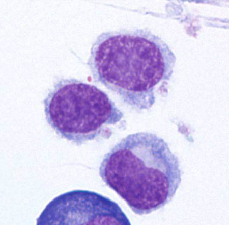
Avaliação pré-tratamento
Os seguintes exames podem orientar o tratamento e devem ser solicitados antes da farmacoterapia ser iniciada:[37]Hoffman MA. Clinical presentations and complications of hairy cell leukemia. Hematol Oncol Clin North Am. 2006 Oct;20(5):1065-73.
http://www.ncbi.nlm.nih.gov/pubmed/16990107?tool=bestpractice.com
Perfil metabólico completo (incluindo ureia, creatinina sérica, eletrólitos, albumina sérica e testes da função hepática [TFHs])
Lactato desidrogenase (LDH) sérica
Sorologia viral para hepatite B e C
Outros exames a serem solicitados
Vários testes podem ser valiosos em circunstâncias específicas.
análise molecular
Pode ser considerada para detectar a mutação BRAF V600E (se houver incerteza diagnóstica após imunofenotipagem) e rearranjos IGHV4-34.[7]Parry-Jones N, Joshi A, Forconi F, et al. Guideline for diagnosis and management of hairy cell leukaemia (HCL) and hairy cell variant (HCL-V). Br J Haematol. 2020 Dec;191(5):730-7.
https://www.doi.org/10.1111/bjh.17055
http://www.ncbi.nlm.nih.gov/pubmed/33053222?tool=bestpractice.com
[34]National Comprehensive Cancer Network. NCCN clinical practice guidelines in oncology: hairy cell leukemia [internet publication].
https://www.nccn.org/professionals/physician_gls/default.aspx
A mutação BRAF V600E está presente em quase todos os pacientes com LCP clássica, mas ausente na LCP-V/SBLPN.[27]Troussard X, Maître E, Paillassa J. Hairy cell leukemia 2024: update on diagnosis, risk-stratification, and treatment - annual updates in hematological malignancies. Am J Hematol. 2024 Apr;99(4):679-96.
https://onlinelibrary.wiley.com/doi/10.1002/ajh.27240
http://www.ncbi.nlm.nih.gov/pubmed/38440808?tool=bestpractice.com
Pacientes com LCP com mutação IGHV4-34 não respondem bem aos tratamentos padrão para a LCP e têm um prognóstico desfavorável. A mutação BRAF V600E geralmente está ausente nesses pacientes.[47]Xi L, Arons E, Navarro W, et al. Both variant and IGHV4-34-expressing hairy cell leukemia lack the BRAF V600E mutation. Blood. 2012 Apr 5;119(14):3330-2.
https://www.doi.org/10.1182/blood-2011-09-379339
http://www.ncbi.nlm.nih.gov/pubmed/22210875?tool=bestpractice.com
Exames de imagem
Não é necessário exame de imagem, exceto em formas mais leves da doença, nas quais tomografias computadorizadas (TC) do tórax, abdome e pelve podem ser usadas para detectar organomegalia e adenopatia leves.[37]Hoffman MA. Clinical presentations and complications of hairy cell leukemia. Hematol Oncol Clin North Am. 2006 Oct;20(5):1065-73.
http://www.ncbi.nlm.nih.gov/pubmed/16990107?tool=bestpractice.com
[34]National Comprehensive Cancer Network. NCCN clinical practice guidelines in oncology: hairy cell leukemia [internet publication].
https://www.nccn.org/professionals/physician_gls/default.aspx
[48]Hakimian D, Tallman MS, Hogan DK, et al. Prospective evaluation of internal adenopathy in a cohort of 43 patients with hairy cell leukemia. J Clin Oncol. 1994 Feb;12(2):268-72.
http://www.ncbi.nlm.nih.gov/pubmed/7906724?tool=bestpractice.com
A TC pode detectar adenopatia interna em aproximadamente 15% dos pacientes.[48]Hakimian D, Tallman MS, Hogan DK, et al. Prospective evaluation of internal adenopathy in a cohort of 43 patients with hairy cell leukemia. J Clin Oncol. 1994 Feb;12(2):268-72.
http://www.ncbi.nlm.nih.gov/pubmed/7906724?tool=bestpractice.com
Lesões osteolíticas raramente são descritas na LCP e têm sido relatadas como um achado em radiografias, normalmente quando os pacientes se apresentam com dor óssea.[49]Robak P, Jesionek-Kupnicka D, Kupnicki P, et al. Bone lesions in hairy cell leukemia: diagnosis and treatment. Eur J Haematol. 2020 Dec;105(6):682-91.
https://www.doi.org/10.1111/ejh.13505
http://www.ncbi.nlm.nih.gov/pubmed/32757401?tool=bestpractice.com