Images and videos
Images


Evaluation of aphasia
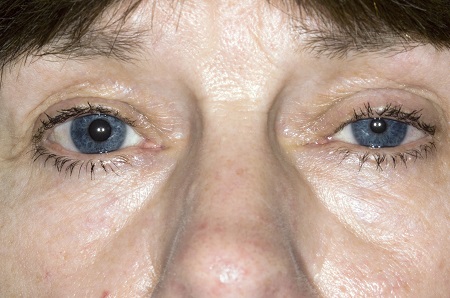
Ptosis and miosis affecting the patient's left eye.
Reproduced with permission from science photo library.
See this image in context in the following section/s:

Evaluation of aphasia
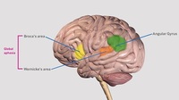
Broca’s area, Wernicke’s area and the angular gyrus.
Created by the BMJ Knowledge Centre.
See this image in context in the following section/s:
Evaluation of aphasia
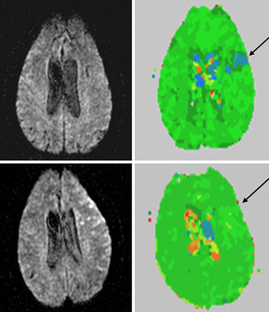
Diffusion-weighted image (top left) and perfusion-weighted image (top right) at day 1 of stroke; arrow points to area of hypoperfusion (blue). Lower panel shows corresponding views at day 2
From the collection of Dr Argye E. Hillis; used with permission
See this image in context in the following section/s:

Evaluation of aphasia
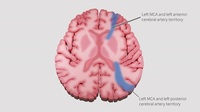
Watershed areas between the anterior, middle and posterior cerebral artery territories.
Created by the BMJ Knowledge Centre.
See this image in context in the following section/s:
Videos
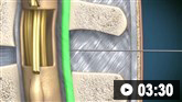 Diagnostic lumbar puncture in adults: animated demonstration
How to perform a diagnostic lumbar puncture in adults. Includes a discussion of patient positioning, choice of needle, and measurement of opening and closing pressure.
Use of this content is subject to our disclaimer