Images and videos
Images


Graft versus host disease
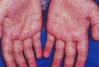
Acute GVHD of the skin (grade I)
Courtesy of Dr John Levine, Professor, Blood and Marrow Transplantation Program, University of Michigan; used with permission
See this image in context in the following section/s:

Graft versus host disease
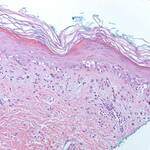
Histology of skin GVHD (high power): Vacuolar interface dermatitis with rare necrotic keratinocytes (200x, hematoxylin and eosin)
Courtesy of Dr Lori Lowe, Professor, Dermatopathology, University of Michigan; used with permission
See this image in context in the following section/s:

Graft versus host disease
Clinical manifestations of chronic graft versus host disease from the National Institutes of Health
Created by BMJ Knowledge Centre
See this image in context in the following section/s:
Graft versus host disease
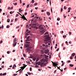
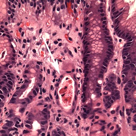
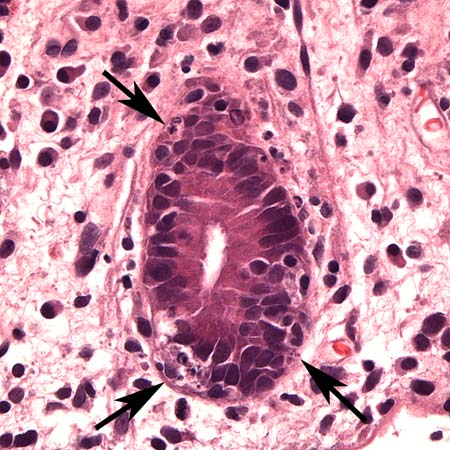
Histology of upper gastrointestinal GVHD (medium-power photomicrograph of the stomach): Dilated gastric gland containing necrotic/apoptotic debris (arrow), typical of GVHD
Courtesy of Dr Joel Greenson, Professor, Pathology, University of Michigan; used with permission
See this image in context in the following section/s:
Graft versus host disease
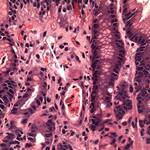
Histology of lower gastrointestinal GVHD (high-power photomicrograph of colon, mild disease): Numerous apoptotic bodies (arrows) indicative of GVHD involving the colon
Courtesy of Dr Joel Greenson, Professor, Pathology, University of Michigan; used with permission
See this image in context in the following section/s:
Graft versus host disease
GVHD pathophysiology
Courtesy of Dr James L.M. Ferrara, Professor, Blood and Marrow Transplantation Program, University of Michigan; used with permission
See this image in context in the following section/s:
Graft versus host disease
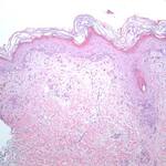
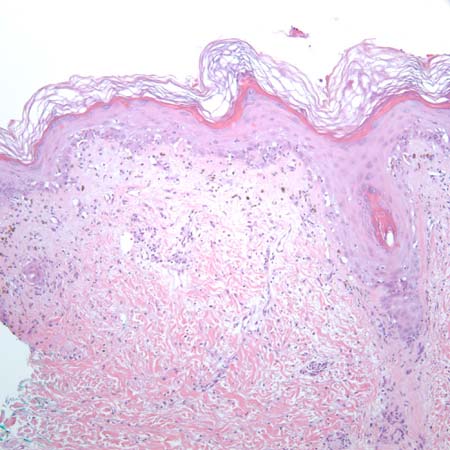
Histology of skin GVHD (low power): Vacuolar interface dermatitis at the dermoepidermal junction with involvement of follicular epithelium (100x, hematoxylin and eosin)
Courtesy of Dr Lori Lowe, Professor, Dermatopathology, University of Michigan; used with permission
See this image in context in the following section/s:

Graft versus host disease
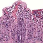
Histology of lower gastrointestinal GVHD (medium-power photomicrograph of colon, severe disease): Almost complete denudation of the mucosa indicative of severe GVHD involving the colon
Courtesy of Dr Joel Greenson, Professor, Pathology, University of Michigan; used with permission
See this image in context in the following section/s:
Use of this content is subject to our disclaimer