Clinical evaluation
Skin examination to identify features associated with eczema should be performed. Affected skin is xerotic (dry) and pruritic, but clinical presentation is variable, depending on age, disease severity, and ethnicity.[47]Kaufman BP, Guttman-Yassky E, Alexis AF. Atopic dermatitis in diverse racial and ethnic groups - variations in epidemiology, genetics, clinical presentation and treatment. Exp Dermatol. 2018 Apr;27(4):340-57.
https://onlinelibrary.wiley.com/doi/10.1111/exd.13514
http://www.ncbi.nlm.nih.gov/pubmed/29457272?tool=bestpractice.com
[48]Silvestre Salvador JF, Romero-Pérez D, Encabo-Durán B. Atopic dermatitis in adults: a diagnostic challenge. J Investig Allergol Clin Immunol. 2017;27(2):78-88.
http://www.jiaci.org/revistas/vol27issue2_1.pdf
http://www.ncbi.nlm.nih.gov/pubmed/28071589?tool=bestpractice.com
[49]Pugliarello S, Cozzi A, Gisondi P, et al. Phenotypes of atopic dermatitis. J Dtsch Dermatol Ges. 2011 Jan;9(1):12-20.
https://onlinelibrary.wiley.com/doi/10.1111/j.1610-0387.2010.07508.x
http://www.ncbi.nlm.nih.gov/pubmed/21054785?tool=bestpractice.com
Acute flares may present with dry skin with erythema and scaling, papules, or vesicles.[49]Pugliarello S, Cozzi A, Gisondi P, et al. Phenotypes of atopic dermatitis. J Dtsch Dermatol Ges. 2011 Jan;9(1):12-20.
https://onlinelibrary.wiley.com/doi/10.1111/j.1610-0387.2010.07508.x
http://www.ncbi.nlm.nih.gov/pubmed/21054785?tool=bestpractice.com
Extensor surfaces, cheeks, and forehead are preferentially affected in infants. Groin and nappy area are usually spared.
In older children and adults, skin flexures are most commonly affected, with hyperpigmentation or hypopigmentation and excoriations from constant scratching.
In chronic eczema, the skin appears thick and lichenified. Other presentations in adults include head and neck dermatitis, hand dermatitis, or prurigo lesions.[48]Silvestre Salvador JF, Romero-Pérez D, Encabo-Durán B. Atopic dermatitis in adults: a diagnostic challenge. J Investig Allergol Clin Immunol. 2017;27(2):78-88.
http://www.jiaci.org/revistas/vol27issue2_1.pdf
http://www.ncbi.nlm.nih.gov/pubmed/28071589?tool=bestpractice.com
Follicular hyperkeratotic papules known as keratosis pilaris (KP) may be present on the extensor surfaces of the upper arms, buttocks, and anterior thighs, and are typically asymptomatic.[49]Pugliarello S, Cozzi A, Gisondi P, et al. Phenotypes of atopic dermatitis. J Dtsch Dermatol Ges. 2011 Jan;9(1):12-20.
https://onlinelibrary.wiley.com/doi/10.1111/j.1610-0387.2010.07508.x
http://www.ncbi.nlm.nih.gov/pubmed/21054785?tool=bestpractice.com
KP are often seen in patients with eczema, but may also be present in the absence of eczema.[Figure caption and citation for the preceding image starts]: Hypopigmentation on the dorsal aspect of the hand in a 12-year-old girl with eczemaFrom the personal collection of A. Hebert, MD; used with permission [Citation ends].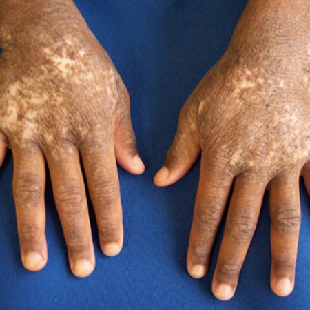 [Figure caption and citation for the preceding image starts]: Papules, lichenification, and hypopigmentation in a child with chronic eczemaFrom the personal collection of A. Hebert, MD; used with permission [Citation ends].
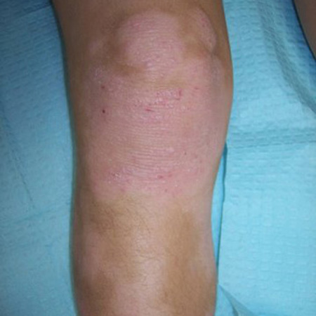 [Figure caption and citation for the preceding image starts]: Hypopigmentation on the flexural skin of the ankles in the same patientFrom the personal collection of A. Hebert, MD; used with permission [Citation ends].
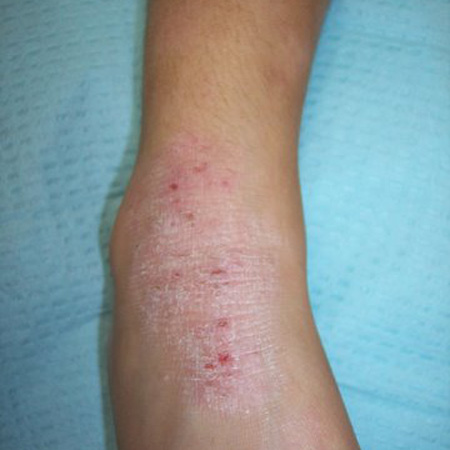
Overlap between acute and chronic eczema may be seen due to the recurrent nature of the disease.[46]National Institute for Health and Care Excellence. Atopic eczema in under 12s: diagnosis and management. Sep 2025 [internet publication].
https://www.nice.org.uk/guidance/cg57
[49]Pugliarello S, Cozzi A, Gisondi P, et al. Phenotypes of atopic dermatitis. J Dtsch Dermatol Ges. 2011 Jan;9(1):12-20.
https://onlinelibrary.wiley.com/doi/10.1111/j.1610-0387.2010.07508.x
http://www.ncbi.nlm.nih.gov/pubmed/21054785?tool=bestpractice.com
The presentation of eczema is considered to be similar amongst ethnic groups. Nuances in the visual appearance of eczema can occur due to differences in pigmentation and distribution of lesions. For example:[46]National Institute for Health and Care Excellence. Atopic eczema in under 12s: diagnosis and management. Sep 2025 [internet publication].
https://www.nice.org.uk/guidance/cg57
[47]Kaufman BP, Guttman-Yassky E, Alexis AF. Atopic dermatitis in diverse racial and ethnic groups - variations in epidemiology, genetics, clinical presentation and treatment. Exp Dermatol. 2018 Apr;27(4):340-57.
https://onlinelibrary.wiley.com/doi/10.1111/exd.13514
http://www.ncbi.nlm.nih.gov/pubmed/29457272?tool=bestpractice.com
More well-demarcated lesions and increased scaling and lichenification are more common in Asian patients
Patients of African descent are less likely to develop flexural dermatitis, but rather present with extensor involvement as a more prominent feature
Discoid or follicular patterns may be more common in Asian, black Caribbean, and black African children.
Consideration of co-existent allergy
There is no pathognomonic marker for the diagnosis of eczema. Food allergy can contribute to disease exacerbation in 30% of children with eczema, with foods such as egg or cows' milk commonly implicated.[50]Eigenmann PA, Sicherer SH, Borkowski TA, et al. Prevalence of IgE-mediated food allergy among children with atopic dermatitis. Pediatrics. 1998 Mar;101(3):E8.
http://www.ncbi.nlm.nih.gov/pubmed/9481027?tool=bestpractice.com
Pollen‐associated food allergy can occur in all ages.[51]European Dermatology Forum. Living EuroGuiDerm guideline for the systemic treatment of atopic eczema. Oct 2023 [internet publication].
https://www.guidelines.edf.one/guidelines/atopic-ezcema
When to consider food allergy testing
Testing for food allergy in patients with eczema should not be considered before intervention with patient education and optimal skin care, including topical corticosteroids, has been initiated.[52]Singh AM, Anvari S, Hauk P, et al. Atopic dermatitis and food allergy: best practices and knowledge gaps - a work group report from the AAAAI Allergic Skin Diseases Committee and Leadership Institute Project. J Allergy Clin Immunol Pract. 2022 Mar;10(3):697-706.
https://www.aaaai.org/Aaaai/media/Media-Library-PDFs/Allergist%20Resources/Statements%20and%20Practice%20Parameters/Atopic-Dermatitis-and-Food-Allergy-Best-Practices-and-Knowledge-Gaps-March-2022.pdf
http://www.ncbi.nlm.nih.gov/pubmed/35101439?tool=bestpractice.com
Patients under 5 years old with moderate to severe eczema, who are unresponsive to initial treatment, should be tested for food allergy using oral food challenge or trial elimination diet.[52]Singh AM, Anvari S, Hauk P, et al. Atopic dermatitis and food allergy: best practices and knowledge gaps - a work group report from the AAAAI Allergic Skin Diseases Committee and Leadership Institute Project. J Allergy Clin Immunol Pract. 2022 Mar;10(3):697-706.
https://www.aaaai.org/Aaaai/media/Media-Library-PDFs/Allergist%20Resources/Statements%20and%20Practice%20Parameters/Atopic-Dermatitis-and-Food-Allergy-Best-Practices-and-Knowledge-Gaps-March-2022.pdf
http://www.ncbi.nlm.nih.gov/pubmed/35101439?tool=bestpractice.com
In the UK, a diagnosis of food allergy should be considered in children with eczema who have reacted to a food with immediate symptoms, or in infants and young children with moderate or severe eczema that is not controlled by optimal management, particularly if associated with gut dysmotility, or with failure to thrive.[46]National Institute for Health and Care Excellence. Atopic eczema in under 12s: diagnosis and management. Sep 2025 [internet publication].
https://www.nice.org.uk/guidance/cg57
Peripheral immunoglobulin E (IgE) levels or skin-prick testing
High sensitivity and low specificity of skin-prick and IgE testing for food allergy can yield false positive results, which may lead to elimination diets that are potentially harmful to patients with eczema.[52]Singh AM, Anvari S, Hauk P, et al. Atopic dermatitis and food allergy: best practices and knowledge gaps - a work group report from the AAAAI Allergic Skin Diseases Committee and Leadership Institute Project. J Allergy Clin Immunol Pract. 2022 Mar;10(3):697-706.
https://www.aaaai.org/Aaaai/media/Media-Library-PDFs/Allergist%20Resources/Statements%20and%20Practice%20Parameters/Atopic-Dermatitis-and-Food-Allergy-Best-Practices-and-Knowledge-Gaps-March-2022.pdf
http://www.ncbi.nlm.nih.gov/pubmed/35101439?tool=bestpractice.com
Effects such as progression to immediate-type allergy, including anaphylactic reactions, have been reported.[52]Singh AM, Anvari S, Hauk P, et al. Atopic dermatitis and food allergy: best practices and knowledge gaps - a work group report from the AAAAI Allergic Skin Diseases Committee and Leadership Institute Project. J Allergy Clin Immunol Pract. 2022 Mar;10(3):697-706.
https://www.aaaai.org/Aaaai/media/Media-Library-PDFs/Allergist%20Resources/Statements%20and%20Practice%20Parameters/Atopic-Dermatitis-and-Food-Allergy-Best-Practices-and-Knowledge-Gaps-March-2022.pdf
http://www.ncbi.nlm.nih.gov/pubmed/35101439?tool=bestpractice.com
[53]Chang A, Robison R, Cai M, et al. Natural history of food-triggered atopic dermatitis and development of immediate reactions in children. J Allergy Clin Immunol Pract. 2016 Mar-Apr;4(2):229-36.e1.
https://www.ncbi.nlm.nih.gov/pmc/articles/PMC4789144
http://www.ncbi.nlm.nih.gov/pubmed/26597013?tool=bestpractice.com
[54]David TJ. Anaphylactic shock during elimination diets for severe atopic eczema. Arch Dis Child. 1984 Oct;59(10):983-6.
https://www.ncbi.nlm.nih.gov/pmc/articles/PMC1628850/pdf/archdisch00729-0083.pdf
http://www.ncbi.nlm.nih.gov/pubmed/6541895?tool=bestpractice.com
[55]Eigenmann PA, Beyer K, Lack G, et al. Are avoidance diets still warranted in children with atopic dermatitis? Pediatr Allergy Immunol. 2020 Jan;31(1):19-26.
http://www.ncbi.nlm.nih.gov/pubmed/31273833?tool=bestpractice.com
Therefore, skin-prick or IgE testing should only be considered for patients who have a history of allergy to food (e.g., exacerbation of eczema after consumption of egg).[52]Singh AM, Anvari S, Hauk P, et al. Atopic dermatitis and food allergy: best practices and knowledge gaps - a work group report from the AAAAI Allergic Skin Diseases Committee and Leadership Institute Project. J Allergy Clin Immunol Pract. 2022 Mar;10(3):697-706.
https://www.aaaai.org/Aaaai/media/Media-Library-PDFs/Allergist%20Resources/Statements%20and%20Practice%20Parameters/Atopic-Dermatitis-and-Food-Allergy-Best-Practices-and-Knowledge-Gaps-March-2022.pdf
http://www.ncbi.nlm.nih.gov/pubmed/35101439?tool=bestpractice.com
[55]Eigenmann PA, Beyer K, Lack G, et al. Are avoidance diets still warranted in children with atopic dermatitis? Pediatr Allergy Immunol. 2020 Jan;31(1):19-26.
http://www.ncbi.nlm.nih.gov/pubmed/31273833?tool=bestpractice.com
[56]Sidbury R, Tom WL, Bergman JN, et al. Guidelines of care for the management of atopic dermatitis: section 4. Prevention of disease flares and use of adjunctive therapies and approaches. J Am Acad Dermatol. 2014 Dec;71(6):1218-33.
https://www.jaad.org/article/S0190-9622(14)01887-8/fulltext
http://www.ncbi.nlm.nih.gov/pubmed/25264237?tool=bestpractice.com
Special consideration should be given to infants with severe eczema, egg allergy, or both, as they have the highest a priori risk for developing peanut allergy.[57]Du Toit G, Roberts G, Sayre PH, et al; LEAP Study Team. Randomized trial of peanut consumption in infants at risk for peanut allergy. N Engl J Med. 2015 Feb 26;372(9):803-13.
https://www.nejm.org/doi/10.1056/NEJMoa1414850
http://www.ncbi.nlm.nih.gov/pubmed/25705822?tool=bestpractice.com
An IgE or skin-prick test is strongly recommended before introducing peanuts into their diet.[52]Singh AM, Anvari S, Hauk P, et al. Atopic dermatitis and food allergy: best practices and knowledge gaps - a work group report from the AAAAI Allergic Skin Diseases Committee and Leadership Institute Project. J Allergy Clin Immunol Pract. 2022 Mar;10(3):697-706.
https://www.aaaai.org/Aaaai/media/Media-Library-PDFs/Allergist%20Resources/Statements%20and%20Practice%20Parameters/Atopic-Dermatitis-and-Food-Allergy-Best-Practices-and-Knowledge-Gaps-March-2022.pdf
http://www.ncbi.nlm.nih.gov/pubmed/35101439?tool=bestpractice.com
The risks and benefits of food testing should be discussed with the patient and their family.
Inhalant allergy
Airborne allergens (including pollen and malassezia yeast) can flare eczema and usually present with exacerbations of disease on the head and neck.[51]European Dermatology Forum. Living EuroGuiDerm guideline for the systemic treatment of atopic eczema. Oct 2023 [internet publication].
https://www.guidelines.edf.one/guidelines/atopic-ezcema
In the UK, a diagnosis of inhalant allergy should be considered in children:[46]National Institute for Health and Care Excellence. Atopic eczema in under 12s: diagnosis and management. Sep 2025 [internet publication].
https://www.nice.org.uk/guidance/cg57
with seasonal flares of eczema
with eczema associated with asthma or allergic rhinitis
aged 3 years or over with eczema on the face, particularly around the eyes.
Patch testing
Allergic contact dermatitis may complicate the clinical course of eczema. Patch testing should be considered for any child or adult whose dermatitis remains difficult to control or presents in a specific location suggestive of an external trigger.[58]Beattie PE, Green C, Lowe G, et al. Which children should we patch test? Clin Exp Dermatol. 2007 Jan;32(1):6-11.
http://www.ncbi.nlm.nih.gov/pubmed/17004990?tool=bestpractice.com
[59]Milingou M, Tagka A, Armenaka M, et al. Patch tests in children: a review of 13 years of experience in comparison with previous data. Pediatr Dermatol. 2010 May-Jun;27(3):255-9.
http://www.ncbi.nlm.nih.gov/pubmed/20609142?tool=bestpractice.com
[60]Pigatto P, Martelli A, Marsili C, et al. Contact dermatitis in children. Ital J Pediatr. 2010 Jan 13;36:2.
https://ijponline.biomedcentral.com/articles/10.1186/1824-7288-36-2
http://www.ncbi.nlm.nih.gov/pubmed/20205907?tool=bestpractice.com
Allergic contact dermatitis may have a more precipitous onset than eczema, and be patchy and irregular in distribution and border. Diagnosis is by patch testing, whereby suspected allergens are placed on unaffected skin of the back for a period of 48 hours. Irritant reactions are evaluated when the patch is removed, and again at subsequent follow-up appointments for delayed reactions. Common contact allergens in patients with eczema include, but are not limited to, nickel, neomycin, fragrance, formaldehyde, and rubber chemicals.[56]Sidbury R, Tom WL, Bergman JN, et al. Guidelines of care for the management of atopic dermatitis: section 4. Prevention of disease flares and use of adjunctive therapies and approaches. J Am Acad Dermatol. 2014 Dec;71(6):1218-33.
https://www.jaad.org/article/S0190-9622(14)01887-8/fulltext
http://www.ncbi.nlm.nih.gov/pubmed/25264237?tool=bestpractice.com
Contact allergy to ingredients in topical therapeutics is known to be increased in people with eczema.[61]Owen JL, Vakharia PP, Silverberg JI. The role and diagnosis of allergic contact dermatitis in patients with atopic dermatitis. Am J Clin Dermatol. 2018 Jun;19(3):293-302.
https://www.ncbi.nlm.nih.gov/pmc/articles/PMC5948135
http://www.ncbi.nlm.nih.gov/pubmed/29305764?tool=bestpractice.com
The discovery of an allergen or allergens in an eczematous child or adult may alleviate further burden of disease and allow better overall responsiveness to appropriate therapy.[62]Jacob SE, Burk CJ, Connelly EA. Patch testing: another steroid-sparing agent to consider in children. Pediatr Dermatol. 2008 Jan-Feb;25(1):81-7.
http://www.ncbi.nlm.nih.gov/pubmed/18304161?tool=bestpractice.com
Treatment of allergic contact dermatitis includes removal of the offending agent and education regarding future avoidance of the contactant.
Skin biopsy
May be considered to differentiate eczema from allergic contact dermatitis, and also from mycosis fungoides or psoriasis.[63]Eichenfield LF, Tom WL, Chamlin SL, et al. Guidelines of care for the management of atopic dermatitis: section 1. Diagnosis and assessment of atopic dermatitis. J Am Acad Dermatol. 2014 Feb;70(2):338-51.
https://www.jaad.org/article/S0190-9622(13)01095-5/fulltext
http://www.ncbi.nlm.nih.gov/pubmed/24290431?tool=bestpractice.com
Used less frequently than skin examination, which considers primary lesions, their distribution, the associated symptomatology, and the skin disease's duration and associations at the time of onset.