Images and videos
Images


Nevi
A common acquired nevus
From the collection of Laurel Schwartz, Thomas Jefferson University
See this image in context in the following section/s:

Nevi
Dermatoscopy of a pigmented Spitz nevus
From the collection of Jason Lee, Thomas Jefferson University
See this image in context in the following section/s:

Nevi
Dermatoscopy of a volar nevus
From the collection of Jason Lee, Thomas Jefferson University
See this image in context in the following section/s:

Nevi
A medium congenital nevus
From the collection of Jason Lee, Thomas Jefferson University
See this image in context in the following section/s:

Nevi
Dermatoscopy of melanoma
From the collection of Laurel Schwartz, Thomas Jefferson University
See this image in context in the following section/s:

Nevi
A common acquired nevus
From the collection of Jason Lee, Thomas Jefferson University
See this image in context in the following section/s:

Nevi
A dysplastic or Clark nevus
From the collection of Jason Lee, Thomas Jefferson University
See this image in context in the following section/s:

Nevi
A persistent or recurrent nevus
From the collection of Jason Lee, Thomas Jefferson University
See this image in context in the following section/s:

Nevi
A pigmented Spitz nevus
From the collection of Jason Lee, Thomas Jefferson University
See this image in context in the following section/s:

Nevi
A Spitz nevus on the ear
From the collection of Jason Lee, Thomas Jefferson University
See this image in context in the following section/s:

Nevi
Dermatoscopy of a blue nevus
From the collection of Jason Lee, Thomas Jefferson University
See this image in context in the following section/s:

Nevi
A volar nevus
From the collection of Jason Lee, Thomas Jefferson University
See this image in context in the following section/s:

Nevi
Dermatoscopy of a dysplastic or Clark nevus
From the collection of Jason Lee, Thomas Jefferson University
See this image in context in the following section/s:

Nevi
Dermatoscopy of a nevus with a reticular pigment network
From the collection of Laurel Schwartz, Thomas Jefferson University
See this image in context in the following section/s:

Nevi
Dermatoscopy of a dysplastic or Clark nevus
From the collection of Jason Lee, Thomas Jefferson University
See this image in context in the following section/s:

Nevi
A giant congenital melanocytic nevus
From the collection of Jason Lee, Thomas Jefferson University
See this image in context in the following section/s:

Nevi
Currently known gene mutations and gene fusions associated with nevi
Derived by Dr JB Lee from: Elder DE et al. WHO Classification of Skin Tumours. 4th ed. International Agency for Research on Cancer; 2018.
See this image in context in the following section/s:

Nevi
A compound acquired melanocytic nevus
From the collection of Laurel Schwartz, Thomas Jefferson University
See this image in context in the following section/s:

Nevi
A dysplastic or Clark nevus
From the collection of Jason Lee, Thomas Jefferson University
See this image in context in the following section/s:

Nevi
A blue nevus
From the collection of Jason Lee, Thomas Jefferson University
See this image in context in the following section/s:

Nevi
A halo nevus
From the collection of Jason Lee, Thomas Jefferson University
See this image in context in the following section/s:

Nevi
Dermatoscopy of a cobblestone pattern with a peripheral pigment network
From the collection of Jason B. Lee, Thomas Jefferson University
See this image in context in the following section/s:

Nevi
A nevus spilus
From the collection of Jason Lee, Thomas Jefferson University
See this image in context in the following section/s:

Nevi
A blue nevus
From the collection of Jason Lee, Thomas Jefferson University
See this image in context in the following section/s:

Nevi
A dysplastic or Clark nevus
From the collection of Jason Lee, Thomas Jefferson University
See this image in context in the following section/s:

Nevi
A small congenital melanocytic nevus
From the collection of Jason Lee, Thomas Jefferson University
See this image in context in the following section/s:

Nevi
Dermatoscopy of a dysplastic or Clark nevus
From the collection of Jason Lee, Thomas Jefferson University
See this image in context in the following section/s:
Videos
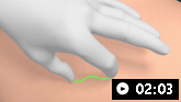 Intradermal injection animation demonstration
Demonstration of injection techniques used to administer local anesthetic, for allergy skin testing, and for tuberculin skin testing.
Use of this content is subject to our disclaimer