Urgent considerations
See Differentials for more details
Non-Hodgkin's lymphoma
The presenting symptoms of non-Hodgkin's lymphoma (NHL) are diverse. People may present with persistent lymphadenopathy, which may occur in association with B symptoms (fever, night sweats, and/or weight loss). NHLs are a heterogenous group of malignancies; the aggressive forms can present acutely.
Examination may reveal localised or generalised lymphadenopathy, hepatosplenomegaly, or skin involvement. Initial investigations should include complete blood count, comprehensive metabolic panel, lactate dehydrogenase, and HIV and hepatitis serologies. The patient should be referred urgently for a hematology consult. An excisional lymph node biopsy is recommended to establish the diagnosis.[24]
Hodgkin's lymphoma
Hodgkin's lymphoma may present with painless cervical or supraclavicular lymphadenopathy, often in association with B symptoms (fever, night sweats and/or weight loss). Pain can occur at sites of lymphadenopathy following alcohol ingestion, likely due to the vasodilatory effect of alcohol within the lymph node capsule. Examination may reveal localised or generalised lymphadenopathy. Initial investigations should include complete blood count. The patient should be referred urgently for a haematology consult. A lymph node excision biopsy establishes the diagnosis.
Superior vena cava syndrome
If lymphadenopathy is associated with regional symptoms such as chest tightness, dysphagia, shortness of breath, or facial swelling indicative of mediastinal disease, a CT scan of the chest should be performed to exclude a mass and/or obstruction of the superior vena cava.
Occlusion or compression of the superior vena cava (SVC) by a mass can result in superior vena cava syndrome.[25] Venous return from the head, thorax, and upper extremities to the right atrium is interrupted. The increased venous pressure results in oedema of the head, neck, and arms. Patients often have symptoms of cyanosis, plethora, and distended vasculature. Prominent collateral veins covering the anterior chest wall may be visible. Bending forwards usually worsens venous engorgement and is a helpful clinical sign. Laryngeal oedema, cyanosis, papilloedema, mental changes, stupor, and coma may occur with severe obstruction.[Figure caption and citation for the preceding image starts]: Prominent collateral vein on the anterior chest and abdominal wall in a patient with superior vena cava syndromeFrom the collection of E Kempny [Citation ends].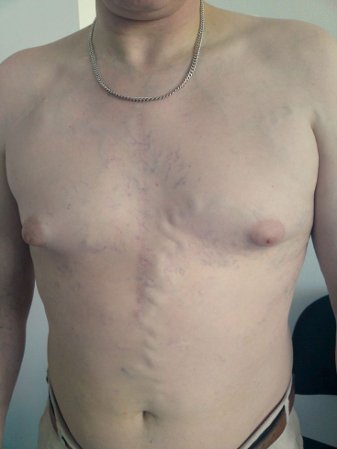
SVC syndrome is a clinical diagnosis and requires a high degree of suspicion. An intrathoracic malignancy is responsible in the majority of cases; non-small cell lung cancer is the most common cause, followed by small cell lung cancer, and non-Hodgkin's lymphoma.
Concern for airway obstruction at presentation necessitates immediate intervention. Treatment consists of securing the airway and relief of laryngeal or cerebral oedema via a combination of corticosteroids and radiation therapy, or percutaneous stenting.
If the patient does not have a known diagnosis of malignancy, then biopsy is required once the patient is stable in order to implement effective therapy. If no peripheral lymph nodes can be sampled, mediastinal lymph nodes can be sampled under the guidance of endoscopic ultrasound, endobronchial ultrasound, CT, or mediastinoscopy.[26]
Use of this content is subject to our disclaimer