Recommendations
Key Recommendations
Have a high index of suspicion; make the diagnosis at the earliest opportunity and have a low threshold for referral for immediate surgical debridement.
Necrotising fasciitis is a life-threatening and time-critical surgical emergency.[3][5]
Discuss the patient early with the critical care team.
Always suspect necrotising fasciitis in a patient with a rapidly progressing soft-tissue infection and any of the following:[3]
Severe pain (disproportionate to the clinical findings) or anaesthesia in the local area.[1][2][3][16][19][20][34][35]
Oedema that extends beyond the erythema.[3]
Systemic signs of infection.[1][3][16][35][36] Sepsis and multi-organ failure may be present.[3][37] Think ' Could this be sepsis?' based on acute deterioration in an adult patient in whom there is clinical evidence or strong suspicion of infection.[38][39][40] See Sepsis in adults.
Practical tip
Think 'Could this be sepsis?' based on acute deterioration in an adult patient in whom there is clinical evidence or strong suspicion of infection.[38][39][40]
Use a systematic approach, alongside your clinical judgement, for assessment; urgently consult a senior clinical decision-maker (e.g., ST4 level doctor in the UK) if you suspect sepsis.[38][40][41][42]
Refer to local guidelines for the recommended approach at your institution for assessment and management of the patient with suspected sepsis.
If you suspect necrotising fasciitis, immediately refer the patient to the surgical team; do not wait for the results of investigations before referral.[2][3] Necrotising fasciitis is a clinical diagnosis. However, investigations can support the diagnosis if this is unclear.[3]
Be aware that the diagnosis can be easily missed because the patient may present early in the disease process with non-specific signs and symptoms.[5]
This topic covers the diagnosis and management of necrotising fasciitis in adults only.
Always suspect necrotising fasciitis in a patient with a rapidly progressing soft-tissue infection and any of the following:[3]
Severe pain (disproportionate to the clinical findings) or anaesthesia over the site of infection[1][2][3][16][19][20][34][35]
Oedema and erythema; oedema will typically extend beyond the erythema[3]
Other possible symptoms of necrotising fasciitis include lightheadedness, palpitations, and nausea or vomiting, or delirium.
Practical tip
Many cases of necrotising fasciitis begin as cellulitis or are misdiagnosed as cellulitis; the patient may present without systemic signs of infection.[5][3] The early differential diagnosis between cellulitis and a necrotising infection that requires prompt surgical intervention may be difficult.[3] See Differentials and Cellulitis and erysipelas.
Be aware that necrotising fasciitis is a clinical diagnosis with signs and symptoms that change rapidly over time.[3][5] Early recognition is critical because necrotising fasciitis is a life-threatening surgical emergency.
Have a high index of suspicion; the diagnosis can be easily missed because the patient may present early in the disease process with non-specific signs and symptoms.[5]
If you suspect necrotising fasciitis, refer the patient immediately for surgical debridement and discuss the patient early with the critical care team; do not wait for the results of investigations.
Take a detailed history; specifically ask about risk factors, including:[1][2][16][24][30][43]
Preceding skin lesions or breakdown
Trauma, surgery
Necrotising fasciitis in the context of recent abdominal surgery or in the groin is most likely to be polymicrobial
Immunosuppression due to chronic illness (e.g., diabetes mellitus, alcohol dependence)
Intravenous drug use
Chickenpox
Herpes zoster
Hospitalisation.
Be aware that the inciting insult may be minor (e.g., an insect bite) and/or not recalled by the patient.[1][26]
Exposure history may occasionally be helpful (e.g., freshwater exposure associated with Aeromonas hydrophila, saltwater exposure or consumption of raw oysters associated with Vibrio vulnificus); however, initial selection of empirical antibiotics should be broad and not guided solely by historical exposures.[9][26]
Assess the patient for systemic signs of infection such as tachycardia, tachypnoea, and hypotension, and toxic shock syndrome (caused by infection with group A streptococcus).[1][2][3][16][35] See Toxic shock syndrome.
Bear in mind that many patients present without systemic signs of infection.[5]
Sepsis and multi-organ failure may also be present.[3][37] Think ' Could this be sepsis?' based on acute deterioration in an adult patient in whom there is clinical evidence or strong suspicion of infection.[38][39][40] See Sepsis in adults.
More info: Sepsis
Think 'Could this be sepsis?' based on acute deterioration in an adult patient in whom there is clinical evidence or strong suspicion of infection.[38][39][40]
The patient may present with non-specific or non-localised symptoms (e.g., acutely unwell with a normal temperature) or there may be severe signs with evidence of multi-organ dysfunction and shock.[38][39][40]
Remember that sepsis represents the severe, life-threatening end of infection.[44]
Necrotising fasciitis is a rapidly progressive disease that can quickly lead to overwhelming sepsis and death; mortality in patients who develop shock and end-organ damage approaches 50% to 70%.[2]
Use a systematic approach (e.g., the National Early Warning Score 2 [NEWS2]), alongside your clinical judgement, to assess the risk of deterioration due to sepsis.[38][39][41][45] Consult local guidelines for the recommended approach at your institution.
Arrange urgent review by a senior clinical decision-maker (e.g., ST4 level doctor in the UK) if you suspect sepsis:[42]
Within 30 minutes for a patient who is critically ill (e.g., NEWS2 score of 7 or more, evidence of septic shock, or other significant clinical concerns)
Within 1 hour for a patient who is severely ill (e.g., NEWS2 score of 5 or 6).
Follow your local protocol for investigation and treatment of all patients with suspected sepsis, or those at risk. Start treatment promptly. Determine urgency of treatment according to likelihood of infection and severity of illness, or according to your local protocol.[42][45]
In the community: refer for emergency medical care in hospital (usually by blue-light ambulance in the UK) any patient who is acutely ill with a suspected infection and is:[40]
Deemed to be at high risk of deterioration due to organ dysfunction (as measured by risk stratification)
At risk of neutropenic sepsis.
If you suspect sepsis due to necrotising fasciitis, immediately refer the patient to the surgical team for inspection, exploration, and debridement of infected tissue.[2][3][5][42]
See Sepsis in adults.
Features of necrotising fasciitis that distinguish it from cellulitis include:
Severe pain or anaesthesia over the site of infection[1][2][3][16][19][20][34][35]
The pain experienced with necrotising fasciitis may be disproportionate to the visible skin changes[35]
Skin changes to the area overlying the infection such as crepitus, vesicles, bullae, greyish discoloration, or oedema extending beyond erythema
However, be aware that the patient can present with normal overlying skin, and that skin changes overlying group A streptococcal necrotising fasciitis are a late sign
Subtle skin changes such as leakage of fluid and oedema precede the overt skin changes of blistering and redness.
The extremities are the most common site for necrotising fasciitis.
About half of cases occur in the extremities, with the remainder affecting the perineum, trunk, or head and neck.[1][3][4][5][16][19][20]
The most common site of group A streptococcal necrotising fasciitis is the thigh. Necrotising fasciitis of a limb, especially the arm, is more likely to be due to group A streptococci than a polymicrobial infection.
Some cases of necrotising fasciitis may have associated myositis due to contiguous spread. This is more common in group A streptococcal than polymicrobial infections.
If you suspect necrotising fasciitis clinically, immediately refer the patient for inspection, exploration, and debridement of infected tissue.[5][46]
The ‘finger test’ is a surgical method that can be performed under local anaesthesia at the bedside for the diagnosis of necrotising fasciitis.[3] It involves making a 2 cm incision down to the deep fascia. Findings that suggest necrotising fasciitis following incision include:[3]
Minimal resistance to finger dissection (a ‘positive’ finger test)
Absence of bleeding
Presence of necrotic tissue
Murky or greyish ‘dishwater’ fluid.
Definitive bacteriological diagnosis is best made from tissue specimens obtained from surgical debridement.[2] Gram staining of clinically affected tissue may provide an early indication of the causative organism(s). For example, small chains of gram-positive cocci suggest a streptococcal infection, whereas clumps of large cocci suggest Staphylococcus aureus.
Early frozen-section soft-tissue biopsy can provide a definitive diagnosis and may be used if the diagnosis is unclear clinically or radiologically.[3] However, frozen-section soft-tissue biopsy requires specialist pathology expertise, takes time to perform, and is not widely available in all regions, including in the UK.[3]
Necrotising fasciitis is classified according to the underlying pathogen as type I or II – see Classification.[5]
[Figure caption and citation for the preceding image starts]: Late signs of necrotising fasciitis with extensive cellulitis, induration, skin necrosis, and formation of haemorrhagic bullaeFrom: Hasham S, Matteucci P, Stanley PRW, et al. Necrotising fasciitis. BMJ. 2005 Apr 9;330(7495):830-3 [Citation ends]. [Figure caption and citation for the preceding image starts]: Necrotising fasciitis on the right abdomen of a 2-year old girl following varicella infectionFrom: de Benedictis FM, Osimani P. Necrotising fasciitis complicating varicella. BMJ Case Rep. 2009;2009:bcr2008141994 [Citation ends].
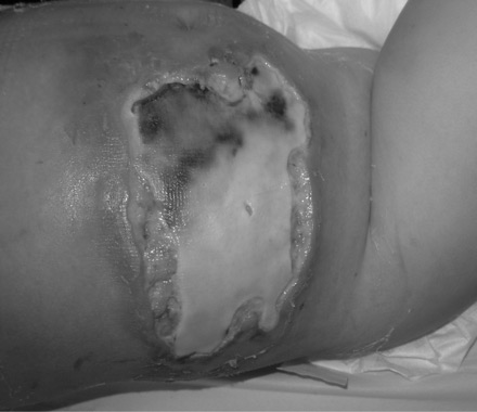 [Figure caption and citation for the preceding image starts]: Split thickness skin grafting after surgical debridementFrom: Hasham S, Matteucci P, Stanley PRW, et al. Necrotising fasciitis. BMJ. 2005 Apr 9;330(7495):830-3 [Citation ends].
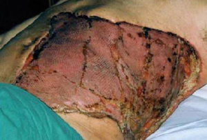
If you suspect necrotising fasciitis, immediately refer the patient to the surgical team; do not wait for the results of investigations before referral.[2][3]
Necrotising fasciitis is a clinical diagnosis. However, investigations can support the diagnosis if this is unclear.[3]
Laboratory tests
Always order:
Blood cultures: obtain these as soon as possible and before starting antibiotics, to help identify the causative organism[3]
Full blood count with white cell differential: may show abnormally high or low white blood cell count with or without a left shift (elevated percentage of polymorphonuclear leukocytes and/or bands)
Urea, electrolytes, and creatinine: urea and creatinine may be elevated due to intracellular volume depletion; serum sodium may be low
C-reactive protein: usually elevated creatine kinase: may be elevated
Liver function tests: may be elevated if there is organ dysfunction due to sepsis
Clotting screen: may show coagulopathy
Blood gas (venous or arterial): lactate is usually elevated. Consider performing an arterial blood gas if you are concerned about respiratory compromise.
Imaging
Imaging may show soft-tissue gas, which is highly suggestive of the diagnosis; imaging may also demonstrate abnormalities in the involved soft tissue.[1][3][4][16]
Seek advice from a radiologist to determine the most appropriate imaging modality for your patient.
Computed tomography (CT) is typically the radiological test of choice.[5]
Do not use a plain x-ray to rule out the diagnosis because x-ray is frequently normal during the early stages; subcutaneous gas may only be present as the disease progresses.[15]
Bedside ultrasound may be performed if the patient is clinically unstable.[3][4] In practice, however, bedside ultrasound is not widely used in all regions (including in the UK).
In one prospective study, ultrasound findings of diffuse thickening of the subcutaneous tissue, accompanied by fluid accumulation greater than 4 mm in depth, had a sensitivity of 88% and a specificity of 93%.[47]
Use a scoring tool to:[3]
Identify patients who are at risk of deterioration
Identify those who need to be managed in a critical care setting
Help distinguish necrotising fasciitis from less severe soft-tissue infections.
Commonly used examples include the following.
The Laboratory Risk Indicator for Necrotising Fasciitis (LRINEC) score[3][4]
LRINEC is based on laboratory parameters and was developed to assist with early discrimination of necrotising fasciitis from less severe skin and soft-tissue infections.[48]
Do not use LRINEC to rule out the diagnosis of necrotising fasciitis.[3] Validation studies have failed to demonstrate sufficient sensitivity or specificity to either diagnose or exclude necrotising fasciitis.[49][50]
Suspect necrotising fasciitis if the patient scores 6 or more; a score of 8 or more is strongly predictive of necrotising fasciitis.[3]
An initial score of greater than 7 is associated with poorer outcomes and higher risk of death in necrotising fasciitis.[51]
Portsmouth Physiological and Operative Severity Score for the enumeration of Mortality and Morbidity (P-POSSUM)
P-POSSUM is a tool that has been validated for estimating an individual patient’s risk of death within 30 days of emergency general surgery, and is based on preoperative and perioperative factors.[52]
If you are using this tool preoperatively, estimate any perioperative factors and update these at the end of surgery.[52]
Transfer the patient to critical care if they have a predicted mortality risk ≥10%.[52]
Use of this content is subject to our disclaimer