Investigations
1st investigations to order
CXR
Test
This test is not diagnostic or very sensitive, but it may be helpful in providing information with regard to the presence or absence of a clinically significant shunt. The CXR can be completely normal for patients with smaller shunts. With more significant shunting it may show cardiomegaly (left atrial and left ventricular enlargement), increased pulmonary markings, left atrial dilation (best seen on the lateral film), and a prominent main pulmonary artery.[33]
Result
cardiomegaly, increased pulmonary markings, prominent main pulmonary artery segment, left atrial dilation
ECG
Test
An ECG is not very sensitive for detecting a patent ductus arteriosus and is used only for corroborative purposes. It may be completely normal. With significant left-sided overload, left ventricular dilation and hypertrophy will result in deep Q waves and tall R waves in leads II, III, aVF, and the left precordial leads (V5 and V6). Significant left atrial enlargement may manifest as widened P waves.[2] These ECG findings are non-specific, as they can be seen with other left-sided shunts such as a ventricular septal defect.
Result
deep Q waves and tall R waves in leads II, III, aVF, V5, and V6. Sometimes widened P waves
echocardiogram
Test
Definitive diagnostic test for a PDA.[33][41] In children the echocardiogram should be performed by a sonographer trained in congenital heart disease in conjunction with a paediatric cardiologist; alternatively, referral to a paediatric cardiologist should be made.[44]
Echocardiography can confirm the presence and size of the ductus as well as the direction of shunting. It is important to determine the direction and velocity of the shunt to understand the ductal resistance and the relative systemic to pulmonary pressure. Left heart chamber dimensions can also be evaluated to determine the significance of the duct.
In the early days of echo, sensitivity was 96% and specificity was 100% compared with angiography.[45] However, in the present era, with echo's improved capabilities, it is accepted as 100% sensitive, with angiography and catheterisation reserved for treatment purposes only.[Figure caption and citation for the preceding image starts]: Echo of a 26-week premature infant demonstrates colour Doppler flow from the systemic circulation (aorta) through the patent ductus arteriosus (PDA) to the left pulmonary arteryNelangi M. Pinto, MD; used with permission [Citation ends].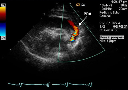
Result
Two-dimensional and/or colour Doppler evidence of a patent ductus arteriosus (PDA). Diastolic forward flow in the pulmonary artery. Left ventricular and/or left atrial enlargement. Diastolic flow reversal in the distal aortic arch
Investigations to consider
cardiac catheterisation and angiography
Test
Not routinely done for diagnostic evaluation of uncomplicated patent ductus arteriosus.
Angiography provides anatomic evidence of shunt. If used diagnostically, it provides evidence of the presence and severity of shunt and allows assessment of any associated pulmonary hypertension.
Role of catheterisation is chiefly therapeutic in facilitating transcatheter occlusion. [Figure caption and citation for the preceding image starts]: Lateral angiogram of 1-year-old child demonstrates flow of contrast from the aorta through a patent ductus arteriosus (PDA) to the pulmonary circulationNelangi M. Pinto, MD; used with permission [Citation ends].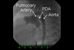
Result
presence of shunt and any co-existing cardiac abnormalities; pulmonary hypertension.
Use of this content is subject to our disclaimer