Differentiation between peri-orbital and orbital cellulitis is imperative as the latter has greater morbidity. Diagnosis of peri-orbital cellulitis is based on history and complete ophthalmological examination. Ophthalmoplegia, proptosis, and visual disturbances are key differentiating factors for orbital cellulitis, and if suspected, a CT or MRI should be ordered to confirm the diagnosis.[20]American Academy of Ophthalmology. Orbital cellulitis. Apr 2024 [internet publication].
https://eyewiki.org/Orbital_Cellulitis
Cultures are usually negative for both, but can be obtained prior to treatment if they are readily accessible. Culture of the nasopharynx and conjunctiva may also be done. However, in most cases, therapy is started empirically.
History
A comprehensive history, along with the pertinent questions about the presenting complaint, should include a history of any recent or current infections (especially of the sinuses, teeth, or ears), styes/chalazions, penetrating injury, and insect bites.[14]Miller JM, Binnicker MJ, Campbell S, et al. Guide to utilization of the microbiology laboratory for diagnosis of infectious diseases: 2024 update by the Infectious Diseases Society of America (IDSA) and the American Society for Microbiology (ASM). Clin Infect Dis. 2024 Mar 5:ciae104.
https://academic.oup.com/cid/advance-article/doi/10.1093/cid/ciae104/7619499
http://www.ncbi.nlm.nih.gov/pubmed/38442248?tool=bestpractice.com
Underlying sinusitis is the overwhelming cause of orbital cellulitis. In children, it frequently accompanies pre-septal cellulitis as well.[1]Robinson A, Beech T, McDermott AL, et al. Investigation and management of adult periorbital or orbital cellulitis. J Laryngol Otol. 2007;121:545-7.
http://www.ncbi.nlm.nih.gov/pubmed/17164026?tool=bestpractice.com
[21]Schramm VL, Myers EN, Kennerdell JS. Orbital complications of acute sinusitis: evaluation, management, and outcome. Otolaryngology. 1978;86:221-230. Any trauma to the eye, direct or indirect (e.g., foreign body in the eye, orbital fracture), should be ruled out. Some patients present with symptoms of this condition after ocular surgery, especially strabismus surgery.[1]Robinson A, Beech T, McDermott AL, et al. Investigation and management of adult periorbital or orbital cellulitis. J Laryngol Otol. 2007;121:545-7.
http://www.ncbi.nlm.nih.gov/pubmed/17164026?tool=bestpractice.com
[9]Powell KR. Orbital and periorbital cellulitis. Pediatr Rev. 1995;16:163-7.
http://www.ncbi.nlm.nih.gov/pubmed/7753730?tool=bestpractice.com
[10]Chandler JR, Langenbrunner DJ, Stevens ER. The pathogenesis of orbital complications in acute sinusitis. Laryngoscope. 1970;80:1414-1428.
http://www.ncbi.nlm.nih.gov/pubmed/5470225?tool=bestpractice.com
[11]Kloek CE, Rubin PA. Role of inflammation in orbital cellulitis. Int Ophthalmol Clin. 2006 Spring;46(2):57-68. Haemophilus influenzae type b (Hib) vaccination status should be noted.
Peri-orbital cellulitis usually presents as redness around the eyelid without significant pain, tenderness, swelling, or fever. Orbital cellulitis usually presents as a severe redness around the eyelid that is swollen and tender to touch. Pain (ocular, ear, or facial), malaise, headache, and fever are more common in orbital cellulitis. Nausea/vomiting and drowsiness may indicate meningeal involvement.[1]Robinson A, Beech T, McDermott AL, et al. Investigation and management of adult periorbital or orbital cellulitis. J Laryngol Otol. 2007;121:545-7.
http://www.ncbi.nlm.nih.gov/pubmed/17164026?tool=bestpractice.com
[9]Powell KR. Orbital and periorbital cellulitis. Pediatr Rev. 1995;16:163-7.
http://www.ncbi.nlm.nih.gov/pubmed/7753730?tool=bestpractice.com
[10]Chandler JR, Langenbrunner DJ, Stevens ER. The pathogenesis of orbital complications in acute sinusitis. Laryngoscope. 1970;80:1414-1428.
http://www.ncbi.nlm.nih.gov/pubmed/5470225?tool=bestpractice.com
[11]Kloek CE, Rubin PA. Role of inflammation in orbital cellulitis. Int Ophthalmol Clin. 2006 Spring;46(2):57-68.[Figure caption and citation for the preceding image starts]: Swollen and red left eyelidFrom the personal collections of H. Jane Kim, MD, and Robert Kersten, MD, UCSF; used with permission [Citation ends].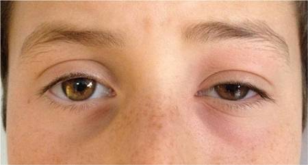
Physical examination
A complete ophthalmological examination should be performed along with a thorough head and neck examination to look for any obvious source of infection such as insect bites to the eye, an infected tooth, or enlarged lymph nodes. Eyelid oedema and erythema are commonly seen in both pre-septal and orbital cellulitis. Visual acuity may be difficult to ascertain in children. It is important to check for a relative afferent pupillary defect, as visual loss due to increased orbital pressure is a real concern in these patients.[1]Robinson A, Beech T, McDermott AL, et al. Investigation and management of adult periorbital or orbital cellulitis. J Laryngol Otol. 2007;121:545-7.
http://www.ncbi.nlm.nih.gov/pubmed/17164026?tool=bestpractice.com
[9]Powell KR. Orbital and periorbital cellulitis. Pediatr Rev. 1995;16:163-7.
http://www.ncbi.nlm.nih.gov/pubmed/7753730?tool=bestpractice.com
[10]Chandler JR, Langenbrunner DJ, Stevens ER. The pathogenesis of orbital complications in acute sinusitis. Laryngoscope. 1970;80:1414-1428.
http://www.ncbi.nlm.nih.gov/pubmed/5470225?tool=bestpractice.com
[11]Kloek CE, Rubin PA. Role of inflammation in orbital cellulitis. Int Ophthalmol Clin. 2006 Spring;46(2):57-68. Intra-ocular pressure may be elevated in orbital cellulitis. This reflects increased orbital pressure due to oedema. Increased orbital pressure can rapidly lead to visual loss and is an indication for urgent intervention. Ophthalmoplegia and proptosis are also key diagnostic factors for orbital cellulitis. Bilateral orbital signs, involvement of the V1 or V2 divisions of the trigeminal nerve, and ophthalmoplegia warrant investigation for cavernous sinus thrombosis. Examine the roof of the mouth and nose for signs of tissue necrosis and a black eschar (late finding) if mucormycosis is suspected (e.g., in immunosuppressed or diabetic patients).
Laboratory investigations
A WBC count, blood cultures and if possible microbiological swab of the conjunctiva, nasopharynx, or any external wound, purulent drainage, or tissue obtained during surgery may be obtained in suspected cases of either peri-orbital or orbital cellulitis.[1]Robinson A, Beech T, McDermott AL, et al. Investigation and management of adult periorbital or orbital cellulitis. J Laryngol Otol. 2007;121:545-7.
http://www.ncbi.nlm.nih.gov/pubmed/17164026?tool=bestpractice.com
[9]Powell KR. Orbital and periorbital cellulitis. Pediatr Rev. 1995;16:163-7.
http://www.ncbi.nlm.nih.gov/pubmed/7753730?tool=bestpractice.com
[10]Chandler JR, Langenbrunner DJ, Stevens ER. The pathogenesis of orbital complications in acute sinusitis. Laryngoscope. 1970;80:1414-1428.
http://www.ncbi.nlm.nih.gov/pubmed/5470225?tool=bestpractice.com
[11]Kloek CE, Rubin PA. Role of inflammation in orbital cellulitis. Int Ophthalmol Clin. 2006 Spring;46(2):57-68.[14]Miller JM, Binnicker MJ, Campbell S, et al. Guide to utilization of the microbiology laboratory for diagnosis of infectious diseases: 2024 update by the Infectious Diseases Society of America (IDSA) and the American Society for Microbiology (ASM). Clin Infect Dis. 2024 Mar 5:ciae104.
https://academic.oup.com/cid/advance-article/doi/10.1093/cid/ciae104/7619499
http://www.ncbi.nlm.nih.gov/pubmed/38442248?tool=bestpractice.com
[15]Leal SM Jr, Rodino KG, Fowler WC, et al. Practical guidance for clinical microbiology laboratories: diagnosis of ocular infections. Clin Microbiol Rev. 2021 Jun 16;34(3):e0007019.
https://www.ncbi.nlm.nih.gov/pmc/articles/PMC8262805
http://www.ncbi.nlm.nih.gov/pubmed/34076493?tool=bestpractice.com
Positive culture rates are between 0% and 33%.[22]Dudin A, Othman A. Acute periorbital swelling: evaluation of management protocol. Pediatr Emerg Care. 1996;12:16-20.
http://www.ncbi.nlm.nih.gov/pubmed/8677172?tool=bestpractice.com
Results are more likely to be positive in children rather than adults.[21]Schramm VL, Myers EN, Kennerdell JS. Orbital complications of acute sinusitis: evaluation, management, and outcome. Otolaryngology. 1978;86:221-230.[23]Patt BS, Manning SC. Blindness resulting from orbital complications of sinusitis. Otolaryngol Head Neck Surg. 1991;104:789-795.
http://www.ncbi.nlm.nih.gov/pubmed/1908969?tool=bestpractice.com
A lumbar puncture is recommended if meningeal signs develop.[12]Swift AC, Charlton G. Sinusitis and the acute orbit in children. J Laryngol Otol. 1990;104:213-216.
http://www.ncbi.nlm.nih.gov/pubmed/2187942?tool=bestpractice.com
[13]Howe L, Jones NS. Guidelines for the management of periorbital cellulitis/abscess. Clin Otolaryngol Allied Sci. 2004;29:725-8.
http://www.ncbi.nlm.nih.gov/pubmed/15533168?tool=bestpractice.com
Imaging
A CT scan with contrast medium is the mainstay diagnostic method for suspected cases of orbital cellulitis, looking for paranasal sinus disease or sub-periosteal abscess.[1]Robinson A, Beech T, McDermott AL, et al. Investigation and management of adult periorbital or orbital cellulitis. J Laryngol Otol. 2007;121:545-7.
http://www.ncbi.nlm.nih.gov/pubmed/17164026?tool=bestpractice.com
[9]Powell KR. Orbital and periorbital cellulitis. Pediatr Rev. 1995;16:163-7.
http://www.ncbi.nlm.nih.gov/pubmed/7753730?tool=bestpractice.com
[10]Chandler JR, Langenbrunner DJ, Stevens ER. The pathogenesis of orbital complications in acute sinusitis. Laryngoscope. 1970;80:1414-1428.
http://www.ncbi.nlm.nih.gov/pubmed/5470225?tool=bestpractice.com
[11]Kloek CE, Rubin PA. Role of inflammation in orbital cellulitis. Int Ophthalmol Clin. 2006 Spring;46(2):57-68.[24]American College of Radiology. ACR appropriateness criteria: orbital imaging and visual loss - child. 2023 [internet publication].
https://acsearch.acr.org/docs/3158173/Narrative
An MRI may be requested to rule out intracranial abscesses and cavernous sinus thrombosis, if clinically indicated.[12]Swift AC, Charlton G. Sinusitis and the acute orbit in children. J Laryngol Otol. 1990;104:213-216.
http://www.ncbi.nlm.nih.gov/pubmed/2187942?tool=bestpractice.com
Orbital ultrasonography can differentiate between pre-septal and orbital infection and may have advantages for some patients as it can be performed at the bedside and does not require sedation; however, it is unable to adequately assess associated sinus or intracranial involvement.[19]Hamed-Azzam S, AlHashash I, Briscoe D, et al. Common orbital infections ~ state of the art ~ Part I. J Ophthalmic Vis Res. 2018 Apr-Jun;13(2):175-82.
https://www.ncbi.nlm.nih.gov/pmc/articles/PMC5905312
http://www.ncbi.nlm.nih.gov/pubmed/29719647?tool=bestpractice.com
[25]Anwar MR, Mahant S, Agbaje-Ojo T, et al. Diagnostic test accuracy of ultrasound for orbital cellulitis: a systematic review. PLoS One. 2023;18(7):e0288011.
https://www.ncbi.nlm.nih.gov/pmc/articles/PMC10325084
http://www.ncbi.nlm.nih.gov/pubmed/37410730?tool=bestpractice.com
[26]Yadalla D, Jayagayathri R, Padmanaban K, et al. Bacterial orbital cellulitis - a review. Indian J Ophthalmol. 2023 Jul;71(7):2687-93.
https://www.ncbi.nlm.nih.gov/pmc/articles/PMC10491050
http://www.ncbi.nlm.nih.gov/pubmed/37417106?tool=bestpractice.com
Referral
Any patient with evidence of peri-orbital oedema and redness accompanied by visual disturbance should be referred to an oculoplastics consultant.[1]Robinson A, Beech T, McDermott AL, et al. Investigation and management of adult periorbital or orbital cellulitis. J Laryngol Otol. 2007;121:545-7.
http://www.ncbi.nlm.nih.gov/pubmed/17164026?tool=bestpractice.com
[9]Powell KR. Orbital and periorbital cellulitis. Pediatr Rev. 1995;16:163-7.
http://www.ncbi.nlm.nih.gov/pubmed/7753730?tool=bestpractice.com
[10]Chandler JR, Langenbrunner DJ, Stevens ER. The pathogenesis of orbital complications in acute sinusitis. Laryngoscope. 1970;80:1414-1428.
http://www.ncbi.nlm.nih.gov/pubmed/5470225?tool=bestpractice.com
[11]Kloek CE, Rubin PA. Role of inflammation in orbital cellulitis. Int Ophthalmol Clin. 2006 Spring;46(2):57-68.