Aetiology
Chronic exposure to UV rays, mostly UVB (290 to 320 nanometres) is a key aetiological factor resulting in damage to keratinocyte DNA and has been implicated in skin carcinogenesis.[23][24]
Animal studies have demonstrated that an increase in telomerase activity prolongs cell life span, delaying apoptosis of cells with mutated DNA. Tumour suppressor gene p53 mutations allow continuous replication and accumulation of mutated keratinocytes with irreparably damaged DNA.[25][26][27][28]
Other factors include chronic exposure to UVA, X-rays, radioisotopes, arsenic, and human papillomavirus (particularly in immunocompromised and organ-transplanted patients, and in patients with epidermodysplasia verruciformis).[29]
Pathophysiology
9UVB radiation causes formation of thymidine dimers in the DNA of keratinocytes. Normally, a repair process, induced by the p53 gene, occurs during a period of cell arrest at G1 phase.[27][30][31]
Cells with excessive damage are normally eliminated through the p53-induced programmed death process (apoptosis). However, chronic UVB exposure targets specifically, and causes mutations of, the p53 gene, located on chromosome 17p132. This mutation leads to the perpetuation and clonal expansion of keratinocytes with damaged DNA, causing formation of AKs.[27][30][31]
As AKs progress to squamous cell carcinoma, the keratinocytes with damaged DNA down-regulate their Fas expression to avoid being killed by T cells, and up-regulate their expression of FasL, to induce apoptosis of T cells.[32]
There is also a suppression of signalling proteins, such as interferon-I-stimulated gene factor 3, which are important regulators of cell proliferation and differentiation. This leads to suppression of interferon-alfa, so it is less able to control the proliferation of keratinocytes.[33][34][35]
Classification
Clinical classification[1][2][3][4]
Regular: 1 or multiple skin-coloured, yellowish, or erythematous rough scaly macules or papules, ill-defined, irregularly shaped, 1 to 5 mm in diameter. [Figure caption and citation for the preceding image starts]: Regular actinic keratosisFrom the collection of the Department of Dermatology and Cutaneous Surgery, University of Miami Miller School of Medicine [Citation ends].
Hyperkeratotic: 1 or multiple skin-coloured, yellowish, or erythematous scaly macules or papules with hyperkeratotic surface, ill-defined, irregularly shaped, 1 to 5 mm in diameter. [Figure caption and citation for the preceding image starts]: Hyperkeratotic actinic keratosisFrom the collection of the Department of Dermatology and Cutaneous Surgery, University of Miami Miller School of Medicine [Citation ends].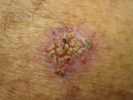
Pigmented: well-defined scaly brown macule or plaque resembling a solar lentigo, and sometimes resembling seborrhoeic keratosis, melanocytic naevus, or an early malignant melanoma (spreading pigmented AKs).[5] A clinical, dermoscopic, and histopathological study has shown that as high as 83% of pigmented AKs were adjacent to or admixed with a separate pigmented lesion, representing a collision between a non-pigmented AK and the pigmented lesion. The most common pigmented lesions were solar lentigo (72%), seborrhoeic keratosis (11%), melanoma in situ (2%), and lichen planus-like keratosis (1%).[6]
Verrucous: skin-coloured, papillomatous, elevated, wart-like papule.
Atrophic: plaques with very mild scale over very thin shiny skin.
Lichen planus-like (lichenoid): violaceous, well-defined papule with fine white lines on the surface.
Actinic cheilitis: scaly red roughness with induration, fissuring, and ulceration of the lower lip to the commissures.[3]
Cutaneous horn: hypertrophic conical-shaped protuberance growing from the surface of the skin; underlying AKs in 19% to 37%.[7][8]
Use of this content is subject to our disclaimer