Images and videos
Images


Evaluation of pruritus
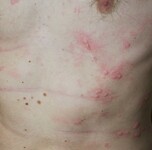
Solar urticaria: photoprovocation upon UVA exposure
From the collection of Adam Reich MD, PhD
See this image in context in the following section/s:

Evaluation of pruritus
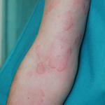
Urticaria: wheals
From the collection of Adam Reich MD, PhD
See this image in context in the following section/s:

Evaluation of pruritus
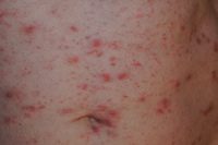
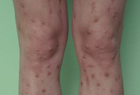
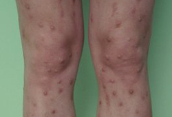
Prurigo nodularis: secondary scratch lesions
From the collection of Adam Reich MD, PhD
See this image in context in the following section/s:

Evaluation of pruritus
Acute urticaria: typical wheals
From the collection of Adam Reich MD, PhD
See this image in context in the following section/s:
Evaluation of pruritus
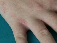
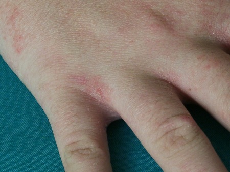
Scabies: typical lesions within interphalangeal areas
From the collection of Adam Reich MD, PhD
See this image in context in the following section/s:

Evaluation of pruritus
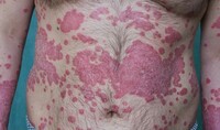
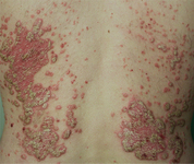
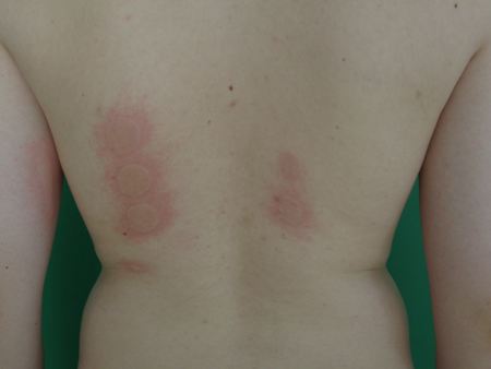
Psoriasis: symmetric plaques on the back covered with thick scales
From the collection of Adam Reich MD, PhD
See this image in context in the following section/s:

Evaluation of pruritus
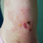
Bullous pemphigoid: tense blisters and erosions on erythematous background
From the collection of Adam Reich MD, PhD
See this image in context in the following section/s:

Evaluation of pruritus
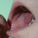
Lichen planus: lacy network traversing the buccal mucosa
From the collection of Adam Reich MD, PhD
See this image in context in the following section/s:

Evaluation of pruritus
Scabies
From the collection of Adam Reich MD, PhD
See this image in context in the following section/s:

Evaluation of pruritus
Atopic dermatitis: white dermographism
From the collection of Adam Reich MD, PhD
See this image in context in the following section/s:

Evaluation of pruritus
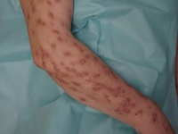
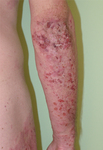
Dermatitis herpetiformis: highly pruritic vesicles, small blisters, and erosions on extensor surfaces of extremities
From the collection of Adam Reich MD, PhD
See this image in context in the following section/s:
Evaluation of pruritus
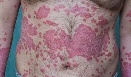
Plaque type psoriasis
From the collection of Adam Reich MD, PhD
See this image in context in the following section/s:
Evaluation of pruritus
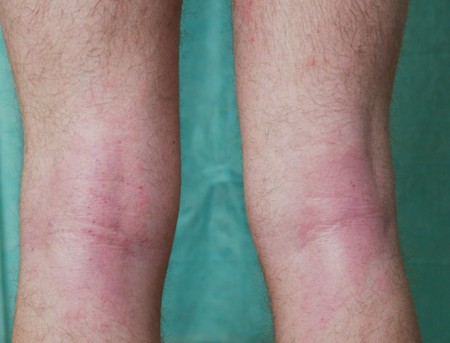
Atopic dermatitis: erythema, excoriations, and lichenification in popliteal area
From the collection of Adam Reich MD, PhD
See this image in context in the following section/s:
Evaluation of pruritus
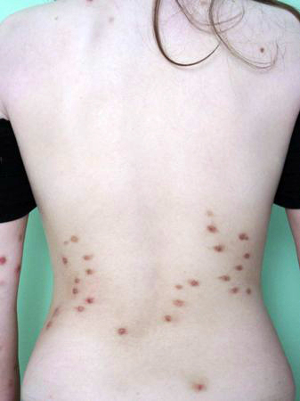
Prurigo nodularis: note sparing of upper part of back, which is difficult to access for scratching
From the collection of Adam Reich MD, PhD
See this image in context in the following section/s:

Evaluation of pruritus
Dermatitis herpetiformis: typical lesions on extensor surface of forearm
From the collection of Adam Reich MD, PhD
See this image in context in the following section/s:

Evaluation of pruritus
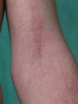
Lichenification due to chronic rubbing in a patient with atopic dermatitis
From the collection of Adam Reich MD, PhD
See this image in context in the following section/s:
Evaluation of pruritus
Prurigo nodularis: a disease with extensive scratch lesions
From the collection of Adam Reich MD, PhD
See this image in context in the following section/s:

Evaluation of pruritus
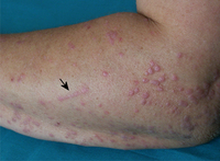
Lichen planus: flat, violaceous papules with visible Köbner phenomenon (arrow)
From the collection of Adam Reich MD, PhD
See this image in context in the following section/s:
Use of this content is subject to our disclaimer