Approach
Diagnosis is made by history and physical examination. Diagnosis is often multi-disciplinary, as the gastrointestinal tract may be involved, or systemic disease may be uncovered in the course of investigation.[18]
[Figure caption and citation for the preceding image starts]: Algorithm for diagnosis of systemic sclerosisFrom the personal collection of Professor J. Pope; used with permission [Citation ends].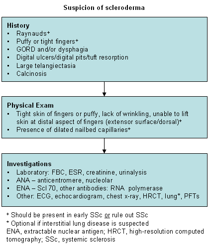
History
At baseline the history and review of systems should include inquiry into:
Silica dust exposure
Duration of Raynaud's phenomenon (RP)
Duration of any skin involvement
Previous carpal tunnel syndrome
Digital ulcers
Any family history of connective tissue diseases, especially scleroderma.
Limited cutaneous systemic sclerosis is characterised by fibrosis of the fingers, rarely with progression of skin fibrosis up to the elbows or knees. It is usually limited to the hands (especially the fingers) or to the feet and distal legs. It does not extend proximally on the upper arms, thighs, or trunk, but it may involve the face and neck. In contrast to this, diffuse cutaneous systemic sclerosis progresses from distal to proximal extremity or to truncal involvement, and may be rapidly progressive; it then may regress with the proximal areas, often softening, but not necessarily with distal skin involvement.
CREST syndrome features (i.e., presence of calcinosis, RP, oesophageal dysmotility, sclerodactyly, and telangiectasia) may be present in both the limited and diffuse types of systemic sclerosis.[Figure caption and citation for the preceding image starts]: Characteristics of limited scleroderma versus diffuse sclerodermaAdapted from LeRoy EC, Black C, Fleischmajer R, et al. Scleroderma (systemic sclerosis): classification, subsets and pathogenesis. J Rheumatol. 1988;15:202-205. [Citation ends].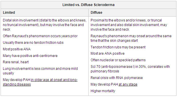
Carpal tunnel or median nerve entrapment is common where there is inflammatory arthritis or tenosynovitis, with numbness sparing the fifth digit. There may also be a history of GORD, but usually dysphagia is a later feature.
Physical examination
On physical examination several items deserve specific notation:
Swollen fingers, with limitation of range of motion (prayer sign) [Figure caption and citation for the preceding image starts]: Prayer sign: fingers cannot touch fully, note also digital tuft resorptionFrom the personal collection of Professor J. Pope; used with permission [Citation ends].

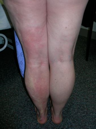
Extent of skin involvement and symmetry (if any) [Figure caption and citation for the preceding image starts]: Linear scleroderma of left leg: note well demarcated changes on left calf, normal skin on lateral sideFrom the personal collection of Professor J. Pope; used with permission [Citation ends].
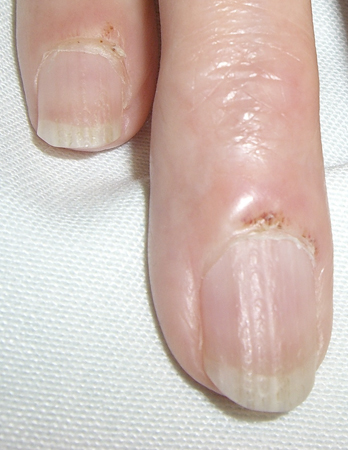
Dilated capillaries at the nailbeds (examined by magnification) [Figure caption and citation for the preceding image starts]: Dilated capillaries at the nailbedsFrom the personal collection of Professor J. Pope; used with permission [Citation ends].
Presence or absence of calcinosis [Figure caption and citation for the preceding image starts]: Calcinosis on second and third fingers in systemic sclerosisFrom the personal collection of Professor J. Pope; used with permission [Citation ends].
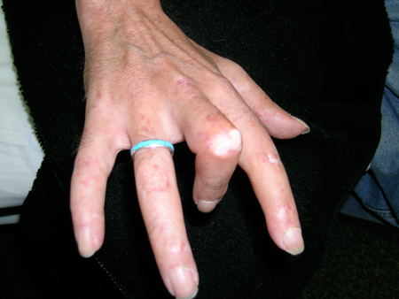
Telangiectasia
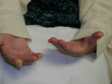
Digital ulcers with pitted scars [Figure caption and citation for the preceding image starts]: Systemic sclerosis with severe digital ulcersFrom the personal collection of Professor J. Pope; used with permission [Citation ends].
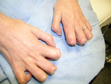
Tendon atrophy (claw hand) [Figure caption and citation for the preceding image starts]: Systemic sclerosis with claw hand deformities (dorsal view)From the personal collection of Professor J. Pope; used with permission [Citation ends].
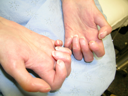
 [Figure caption and citation for the preceding image starts]: Claw hands, palmar view; patient cannot extend fingersFrom the personal collection of Professor J. Pope; used with permission [Citation ends].
Flexion contractures [Figure caption and citation for the preceding image starts]: Multiple telangiectasia, finger flexion deformity, pits on third finger, healed ulcers on second and fourth fingersFrom the personal collection of Professor J. Pope; used with permission [Citation ends].

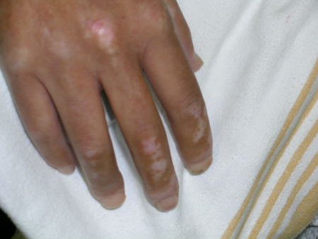
Changes in pigmentation (increased and or decreased pigmentation). [Figure caption and citation for the preceding image starts]: Diffuse cutaneous systemic sclerosis (dsSSc): tight hand and fingers, increased pigmentation, and thumb ulcerFrom the personal collection of Professor J. Pope; used with permission [Citation ends].
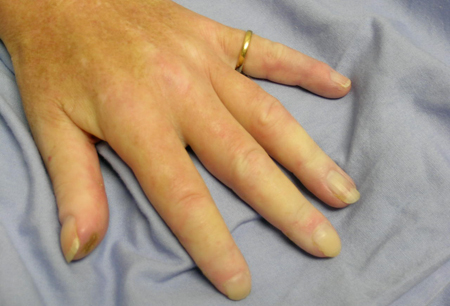 [Figure caption and citation for the preceding image starts]: Sclerodactyly with increased and decreased pigmentationFrom the personal collection of Professor J. Pope; used with permission [Citation ends].
Key features include symmetrical puffy fingers or tight skin, and RP. There may be carpal tunnel findings with positive Tinel's and Phalen's tests due to hand swelling and inflammatory arthritis.
Tinel's test is performed by tapping the median nerve along its course in the wrist. Worsening of the tingling in the fingers is considered positive.
Phalen's test compresses the carpal tunnel in dorsiflexion of the hand with reproduction of the symptoms. Carpal tunnel syndrome is common in the general population. It occurs often in systemic sclerosis when there is swelling of the hands.
Dilated (enlarged) capillaries at the nailbed are visible to the naked eye or with simple magnification (e.g., with an ophthalmoscope). Calcinosis and telangiectasia (well-demarcated large red circles that are blanchable) may not be present early.
Laboratory
A skin biopsy is not needed, and the laboratory testing is mostly confirmatory. It does not make (rule in or rule out) a diagnosis. Some typical findings include:
Full blood count with peripheral fragmentation of red blood cells (associated with systemic disease), but this is rare except when occurring in scleroderma renal crisis
Serum creatinine elevated in the presence of renal crisis (associated with systemic disease)
Serum antinuclear antibodies (may be positive) and extractable nuclear antigens present with antibodies to Scl 70 (anti-topoisomerase I) or RNA polymerase III
Serum erythrocyte sedimentation rate may be elevated and is associated with a worse prognosis.
The detection and measurement of auto-antibodies has a role in diagnosis and prognosis of patients with systemic sclerosis. Certain auto-antibodies have been shown to correlate closely with various clinical and laboratory manifestations of the disease and have certain prognostic value. For example, presence of anti-centromere antibody is associated with a favourable prognosis.[19]
Other investigations
Annual echocardiograms are recommended to screen for pulmonary arterial hypertension (PAH) in systemic sclerosis. Some experts also recommend baseline and annual pulmonary function tests, as a very low diffusing capacity (DLCO) can increase the likelihood of PAH. If both DLCO and forced vital capacity are equally reduced, the possibility of interstitial lung disease (ILD) is increased.
Some experts recommend a baseline high-resolution computed tomography scan of the chest. The scan is repeated if there are new symptoms that might indicate ILD or if there is a clinically significant decline in pulmonary function.[18][20][21][22] Anti-topoisomerase I antibodies have been associated with an increased risk of severe ILD.[23]
In general, oesophageal manometry is at best only contributory to the diagnosis, as dysphagia is quite common. Most patients have abnormal manometry with poorly coordinated peristalsis and diminished strength of contractions.
Use of this content is subject to our disclaimer